
You’re petting your dog one evening and your fingers find something unexpected. A small bump near the lip. A rough cluster on the gum. A raised nodule on the belly that wasn’t there last month. Your mind goes to the worst-case scenario immediately, but before panic sets in, it helps to understand what you’re actually looking at.
Most skin growths on dogs are benign, and dog warts are among the most common. But most does not mean all. The challenge is knowing which growths deserve a calm wait-and-see approach and which ones need a veterinarian’s attention promptly. That distinction is harder to make than it seems, because some dangerous lesions look nearly identical to harmless ones.
CompanAIn was built for moments like this. By consolidating your dog’s health records, documenting skin changes over time, and building a continuously evolving picture of your dog’s overall health, CompanAIn’s agentic AI platform gives your veterinarian the context they need to make confident calls rather than evaluating a mystery bump in isolation.
What Is a Dog Wart?
Clinically, a dog wart is a viral papilloma, a benign growth caused by one of the many strains of canine papillomavirus (CPV). These are not the same virus that causes warts in people. CPV is species-specific and cannot be transmitted to humans or cats, and the human papillomavirus cannot infect dogs.
The virus enters through compromised skin, a small cut, a moist abrasion, or an insect bite. Once inside, it inserts its genetic material into the host cell’s DNA, disrupting normal cell division and driving abnormal proliferation. The result is a visible growth on the skin or mucous membranes.
How Dogs Get Warts
CPV spreads through direct contact with an infected dog or shared objects like food bowls, toys, and bedding. The virus can survive in the environment for weeks, which is why dog parks, daycares, and boarding facilities pose a real transmission risk. After exposure, warts typically take four to six weeks to develop, though a healthy immune system will fight off the virus before any growth appears.
Dogs most likely to develop visible warts are puppies with still-maturing immune systems and older or immunocompromised dogs whose defenses have weakened.
Types of Canine Papillomas
Not all dog warts look alike. Location and appearance often point toward which strain is responsible.
Oral papillomas are the most common presentation. These warts develop on the lips, gums, tongue, and inner cheeks of young dogs, typically under two years of age. They appear as cauliflower-like or fringed growths, grayish-white to flesh-colored, and usually arrive in clusters rather than as a single lesion. They are caused by CPV-1 and tend to resolve within two to three months as the dog builds immunity.
Cutaneous warts appear on the face, eyelids, feet, and lower abdomen. They are considered the second most common skin tumor in dogs under one year of age, and most regress within about three months.
Cutaneous inverted papillomas grow downward into the skin rather than outward, presenting as smooth raised nodules, often on the belly, with a small central pore. Because they lack the cauliflower appearance, they are more easily mistaken for other lesion types.
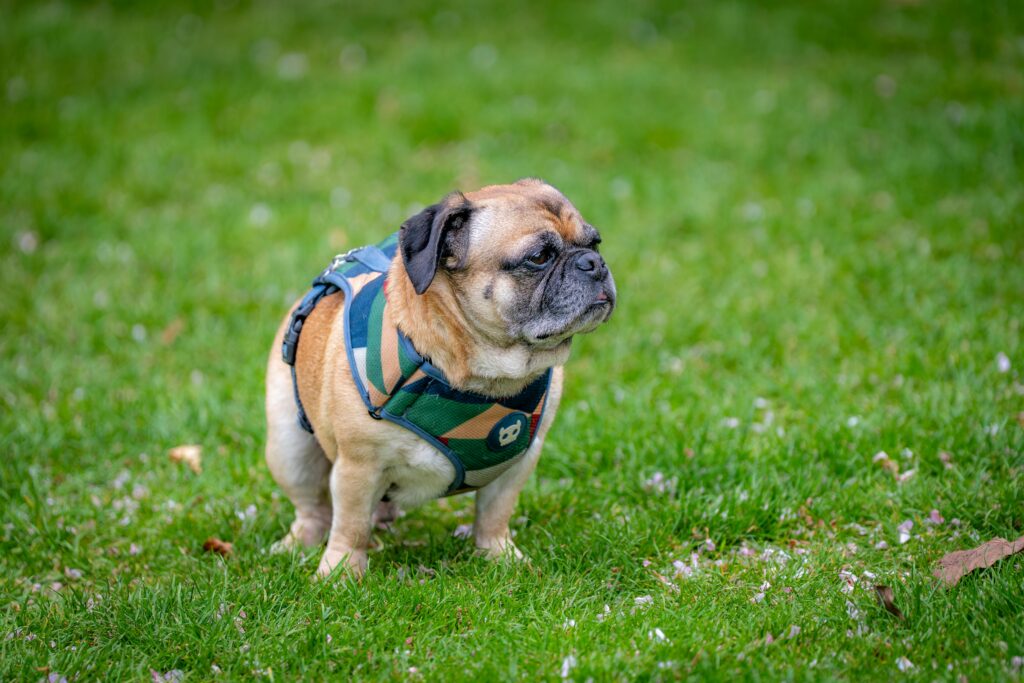
Pigmented plaques are dark, scaly lesions found primarily on the belly and inner legs, more common in Pugs and Miniature Schnauzers. Unlike other papilloma types, they do not reliably regress and carry a higher risk of malignant transformation.
Why a Photo Is Never a Diagnosis
Here is the reality that makes dog warts genuinely complicated: several serious conditions look nearly identical to benign papillomas on visual inspection alone.
Mast cell tumors account for between 7 and 21 percent of all canine skin tumors according to VCA Animal Hospitals, making them the most common malignant skin tumor in dogs. They have earned the nickname “the great pretenders” because they convincingly mimic warts, insect bites, and benign skin tags. A mast cell tumor can appear as a small, firm raised lump or a soft nodule just beneath the skin. It can stay the same size for months or grow and shrink unpredictably. There is no reliable way to distinguish one from a benign papilloma by looking at it.
Squamous cell carcinoma can produce raised, wart-like masses that are firm to the touch. Research from NC State’s College of Veterinary Medicine also notes that certain papillomavirus strains have been linked to squamous cell carcinoma development, meaning the line between benign papilloma and malignant transformation is not always clean.
Sebaceous adenomas are benign growths from oil glands that closely resemble papillomas in older dogs. Harmless, but indistinguishable without testing.
A fine needle aspirate (FNA) or biopsy, evaluated by a veterinary pathologist, is the only way to reliably know what a growth actually is. This applies especially to any lesion that is new, growing, changing in appearance, or appearing on a middle-aged or older dog.
What AI Photo Analysis Does
Visual inspection alone has a well-documented reliability problem in veterinary dermatology. Computer vision applied to skin lesions works by analyzing border regularity, texture, color distribution, and morphology, then comparing those properties against patterns derived from large annotated datasets.
Research published in Veterinary Dermatology demonstrated that convolutional neural network models trained on images of canine paw lesions could distinguish between healthy tissue, inflammatory disease, and neoplasia in real time, with a mean average precision of 0.95. Visual features that appear similar to the human eye carry measurable differences in their underlying properties that AI pattern recognition can detect.
Where Serial Documentation Changes the Picture
A single photo analyzed in isolation answers a limited question. What AI-assisted longitudinal analysis adds is comparison across time, and for skin lesions specifically, that comparison is where the most clinically meaningful information lives.
The visual properties that matter most for distinguishing benign from concerning lesions are dynamic, not static:
- Border regularity that was smooth six months ago and is now irregular
- A lesion that has shifted from uniform to mottled in color
- Diameter that has increased measurably across documented images
- Surface texture that has changed from smooth to ulcerated
None of these changes are visible in a single photograph. They only become apparent when images of the same lesion are analyzed across time against a consistent baseline. Multi-agent AI cross-references each uploaded observation against the documented history of that specific lesion, flagging morphological shifts that neither owners nor veterinarians can reliably detect from memory alone.
This is the distinction between photo analysis as a one-time screening tool and photo analysis as a longitudinal monitoring system. The first tells you what a lesion looks like today. The second tells you whether it looks different from what it looked like before, which is often the more important clinical question.
When Treatment Is Needed
Many true papillomas resolve without any intervention as the immune system suppresses the virus. Oral papillomas typically regress within six to eight weeks; cutaneous lesions usually clear within three months.
Treatment becomes necessary when:
- Growths have been present for more than three to five months without regression
- Warts are bleeding, infected, or ulcerated from scratching
- Growths interfere with eating, walking, or vision
- The dog is on immunosuppressive medications and cannot clear the virus
- Pigmented plaques are present, given their malignant potential
Treatment options include surgical excision, cryotherapy with liquid nitrogen, and topical imiquimod, which stimulates localized immune activity. Antiviral interferon and the antibiotic azithromycin have also been used in severe cases, though results vary.
Immunosuppression is one of the most underappreciated drivers of persistent warts. Cornell University’s College of Veterinary Medicine documented a case where a French Bulldog on a daily corticosteroid-containing allergy medication developed rapidly multiplying skin papillomas. The Cornell team managed the outbreak by gradually reducing the steroid dose rather than pursuing aggressive surgical removal, allowing the dog’s own immune system to take over.
How CompanAIn Supports Skin Lesion Monitoring
A growth noted at a single appointment is just a data point. What veterinarians need to make confident decisions is context: How long has this been there? Has it changed? What medications is this dog on? What does the broader health picture look like?
CompanAIn’s agentic AI platform builds that context. When you upload veterinary records, exam notes, and owner observations through Smart Upload, the platform organizes everything into a structured, filterable Living Health Timeline. A growth noted in passing at one wellness visit does not get lost in old paperwork. It becomes a reference point that informs every future encounter.
Longitudinal Lesion Records
A papilloma that has remained stable for eight months tells a very different story than one that has doubled in size over six weeks. CompanAIn’s Living Memory technology maintains context across years of health history, so when a new growth appears, your veterinarian can immediately see whether anything similar was documented before, whether appearance has changed, and whether any medication adjustments coincide with the timing.
Keeping Risk Factors Visible
Boxers, Pugs, Boston Terriers, and other brachycephalic breeds carry elevated mast cell tumor risk. Dogs on long-term corticosteroids are more vulnerable to persistent papillomavirus infections. Older dogs should have any new growth evaluated promptly, regardless of how innocent it looks.
CompanAIn keeps these risk factors visible at every appointment. Breed predispositions, current medications, and immune status are all part of the picture your veterinarian sees when reviewing the Living Health Timeline, rather than details that need to be reconstructed from memory.
Vet-Ready Summaries
Pet owners often notice changes between appointments but struggle to describe them precisely. CompanAIn’s Vet-Ready AI Summary organizes your dog’s health data into a clinician-grade report your veterinary team can review, so every appointment starts with the full picture rather than a verbal reconstruction of what you can remember.
CompanAIn does not diagnose skin lesions and does not replace the FNA or biopsy that definitively identifies a growth. What it does is ensure your veterinarian has everything needed to make the right call, and so subtle changes are never lost between visits.
Start Building Your Dog's Health Timeline Today
Skin lesions feel urgent the moment you find them and are easy to put off once the initial panic fades. The problem is that the window for simple, effective intervention does not stay open indefinitely.
Contact CompanAIn today to build the Living Health Timeline that keeps every detail of your dog’s health organized and accessible when it matters most. When your veterinarian can see the full picture, they can catch problems early and act with confidence.
Frequently Asked Questions
Are dog warts painful?
Most are not. Oral papillomas occasionally cause mild discomfort when chewing if they are large or numerous, and warts located on the paws can become irritated from friction during walking. Warts that are actively painful, bleeding, or causing the dog to scratch or bite at them consistently warrant veterinary attention regardless of how long they have been present.
Can my dog get warts from the dog park?
Yes. CPV spreads through direct contact with infected dogs and contaminated surfaces including shared water bowls, toys, and agility equipment. The virus can survive in the environment for extended periods, which is why high-traffic dog facilities carry real transmission risks. Dogs with active oral or cutaneous warts should avoid shared spaces until lesions have resolved.
Should I pop or squeeze a dog wart?
No. Manipulating a growth before it has been identified can cause bleeding, introduce infection, and in the case of a mast cell tumor, potentially trigger degranulation and a systemic reaction. Leave any unidentified growth alone until a veterinarian has evaluated it.
How does a vet actually diagnose a skin growth in dogs?
The only definitive method is cytology or histopathology. A fine needle aspirate collects cells from the growth for microscopic evaluation and is minimally invasive. A biopsy removes tissue for more detailed analysis and is recommended when FNA results are inconclusive or when a lesion has features suggesting malignancy. Visual diagnosis alone, including by experienced veterinarians, is not reliable enough for growths that could be mast cell tumors.
What does it mean if my dog's wart changes color?
Color change in any skin lesion is a reason to have it evaluated promptly. Darkening can suggest increased pigmentation or vascular changes associated with growth activity. A lesion shifting from flesh-colored to darker, or developing uneven pigmentation, should not be attributed to normal variation without veterinary confirmation.