
Equine protozoal myeloencephalitis, or EPM, is one of the most frustrating neurologic diseases in horses because it can look like many other problems at first. A horse may seem a little weak behind, start dragging a toe, lose topline on one side, or just feel “off” under saddle before the picture becomes more obvious. EPM is a protozoal infection of the central nervous system, caused most commonly by Sarcocystis neurona and less often by Neospora hughesi. It occurs primarily in the Americas and can affect the brain, brainstem, or spinal cord, which is why the signs vary so much from horse to horse. The signs of EPM can closely resemble those of other neurologic diseases, making diagnosis challenging because symptoms may be similar or even transient.
For owners, the practical challenge is not memorizing every neurologic differential. It is recognizing when asymmetry, weakness, muscle loss, or unexplained incoordination deserves a prompt veterinary workup instead of a wait-and-see approach. EPM cases typically occur as isolated incidents, with rarely more than one horse affected on a given property. That matters because earlier evaluation improves the odds of getting to the right diagnosis and starting appropriate treatment before deficits become entrenched. If you are already trying to separate “monitor closely” from “call the vet now,” CompanAIn’s EquestrAIn guide to horse emergency symptoms is a useful companion for triaging what should not wait.
What equine protozoal myeloencephalitis (EPM) actually is
EPM develops when protozoal organisms enter the horse’s central nervous system and trigger inflammation and tissue damage. The life cycle of these protozoan parasites involves opossums, which are the definitive hosts that shed infective sporocysts in their feces. Horses become infected when they ingest these infective sporocysts through contaminated feed or water. In endemic regions, Sarcocystis neurona is the dominant cause, and infection is associated with ingestion of sporocysts shed by opossums in contaminated feed or water. After ingestion, the sporocysts first enter the horse’s intestinal tract, then, in rare cases, migrate into the bloodstream and cross the blood brain barrier to reach the central nervous system, leading to neurological disease. Exposure is common, but disease is much less common than exposure, which is one reason EPM remains so tricky: a positive blood test alone does not prove that neurologic signs are being caused by EPM. Many clinically normal horses may have antibodies to S. neurona without showing signs of EPM, complicating diagnosis. That distinction is emphasized in the AAEP’s EPM guidelines, the Merck Veterinary Manual’s equine EPM overview, and the ACVIM’s updated consensus statement on EPM.
That same exposure-versus-disease gap is also why owners often feel confused after the first round of testing. Not all infected horses develop clinical signs, so diagnosis relies on a combination of clinical and laboratory findings. EPM is not diagnosed from one clue in isolation. It is diagnosed by putting the neurologic exam, the horse’s history, and targeted testing together.
Common clinical signs of EPM in horses
The hallmark of EPM is an asymmetric neurologic picture. Many horses show weakness, incoordination, or muscle atrophy that is worse on one side than the other. Neurological symptoms such as ataxia, muscle atrophy, and seizures may also be present, reflecting central nervous system involvement. Others may have stumbling, toe dragging, difficulty backing, abnormal tail pull strength, or trouble maintaining balance on circles or slopes. Because any part of the central nervous system can be involved, some horses also develop cranial nerve deficits such as facial nerve paralysis, head tilt, trouble swallowing, or abnormal tongue tone. AAEP’s owner handout, Learn to Recognize the Symptoms of EPM, captures this well: EPM is a “master of disguise,” and signs can range from subtle to severe.
Signs owners commonly notice include:
- asymmetric ataxia or weakness
- dragging a hind toe or scuffing shoes
- unexplained lameness-like gait changes that do not localize cleanly to one limb
- focal muscle wasting, often over the gluteals or topline
- poor coordination going downhill, backing, or turning
- facial nerve paralysis, head tilt, or difficulty swallowing in some cases
- generalized weakness, especially in advanced cases
It is crucial to closely monitor the horse’s symptoms, as recognizing changes in severity can guide timely intervention and help determine the most effective treatment plan.
One reason EPM is so often discussed alongside “mystery performance decline” is that mild cases can look like training issues, soreness, or intermittent lameness before the deficits are unmistakable. That is where longitudinal notes, videos, and lab trends become genuinely useful. CompanAIn’s EquestrAIn article on next-gen equine diagnostics is relevant here because subtle neurologic change is exactly the kind of pattern owners may miss when they rely on memory instead of side-by-side observations over time.
What can look like EPM but is not
EPM is not the only cause of ataxia, weakness, or muscle asymmetry in horses. Cervical vertebral compressive myelopathy, equine motor neuron disease, trauma, equine herpesvirus myeloencephalopathy, viral encephalitides, peripheral nerve injury, lameness, and other inflammatory or degenerative neurologic diseases can overlap with it clinically. Diagnostic tests such as blood tests and spinal taps are often used to help differentiate EPM from other diseases with similar symptoms. That is one reason veterinarians do not diagnose EPM from gait video or symptoms alone. The ACVIM consensus statement and Merck both stress that EPM remains a diagnosis reached in context, not a shortcut diagnosis for every horse with neurologic signs.
For owners, the actionable point is simple: if the signs are asymmetric, progressive, or neurologic-looking, it is time for a veterinary exam rather than trial-and-error management.
How veterinarians diagnose EPM in the central nervous system
Diagnosing EPM in horses is a complex process, as EPM diagnosis in live horses requires careful evaluation of clinical signs and multiple diagnostic tests to distinguish it from other neurological diseases.
A proper EPM workup starts with the neurologic exam. Your veterinarian is looking for evidence that the problem localizes to the central nervous system, whether deficits are symmetric or asymmetric, whether cranial nerves are involved, and how severe the horse’s ataxia and weakness really are. Only after that does EPM-specific testing become meaningful.
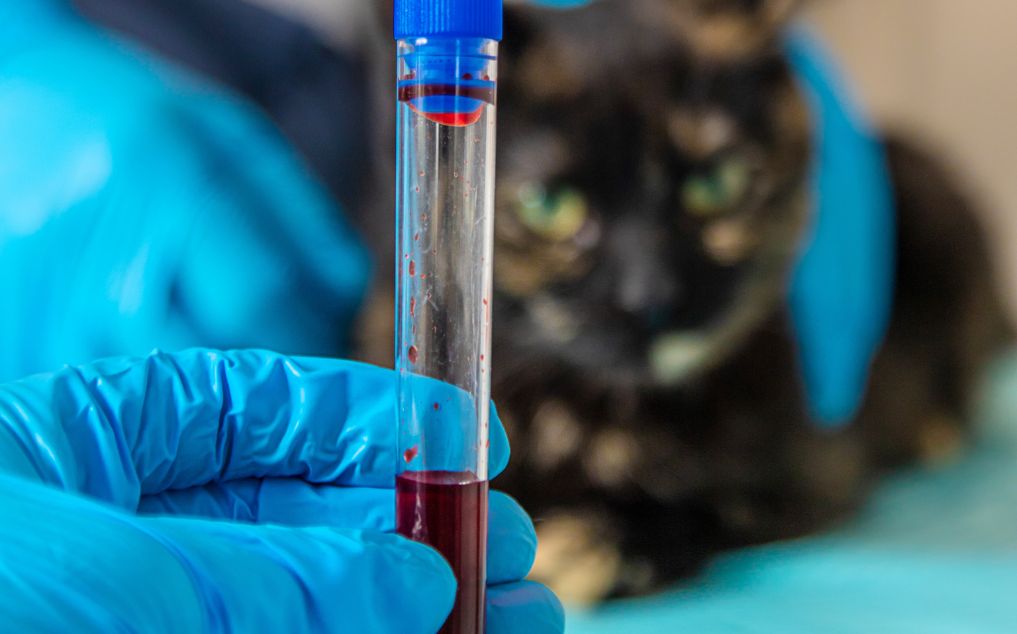
Testing may include serum and cerebrospinal fluid antibody testing, often interpreted as a serum:CSF titer ratio rather than as a simple positive-or-negative exposure result. For accurate diagnosing EPM, analysis of both the horse’s blood and spinal fluid is important, as this helps confirm the diagnosis and distinguish EPM from other neurological conditions. That matters because many healthy horses in endemic regions have been exposed to Sarcocystis neurona without having active neurologic disease. The ACVIM consensus statement strongly emphasizes this point, and newer discussions in The Horse also highlight that serum:CSF titer interpretation can be more informative and cost-effective than treating presumptively without that context. The indirect fluorescent antibody test (IFAT) is a sensitive and specific method for detecting antibodies to S. neurona and N. hughesi, offering quantitative results and advantages over other tests in identifying active EPM infection and supporting clinical diagnosis.
In practice, diagnosis often combines:
- neurologic examination and lesion localization
- serum testing for exposure
- CSF testing when indicated
- interpretation of serum:CSF ratios
- identification of active EPM infection through advanced antibody testing and intrathecal antibody production
- exclusion of other neurologic or lameness causes
- response to treatment as part of the full clinical picture, not as the sole proof
If your vet is also looking at CBC, chemistry, inflammatory markers, or infectious-disease screening while sorting through the case, CompanAIn’s EquestrAIn guide on how to read a horse blood test is a useful owner-facing resource for understanding why those supporting data points matter even when bloodwork alone cannot diagnose EPM.
FDA approved treatments for EPM
Treatment is aimed at reducing or eliminating the protozoal infection and controlling secondary inflammation. Commonly used FDA-recognized treatment approaches include ponazuril, diclazuril, or sulfadiazine/pyrimethamine-based protocols, depending on the case and the veterinarian’s treatment plan. Supportive management may also include anti-inflammatory therapy. If there is evidence of brain involvement, additional treatments such as corticosteroids or other anti-inflammatory agents may be considered to reduce neurological inflammation. Nutritional support is important, with vitamin E supplementation often recommended to support nerve health and aid recovery. Supporting the horse’s immune system through balanced nutrition and stress reduction is also crucial during treatment. Physical rehabilitation and careful adjustment of training expectations during recovery are advised. AAEP and Merck both describe treatment as time-sensitive and individualized, while recent veterinary coverage in The Horse highlights ongoing interest in improving protocols and outcomes.
A few practical points matter for owners:
- improvement is often gradual, not immediate
- some horses improve substantially, but not all return fully to prior athletic function
- relapse or incomplete recovery can occur
- the horse’s starting severity influences prognosis
- rehab and safe management after diagnosis are part of the treatment story, not an afterthought
Prognosis: can horses recover from EPM?
Yes, many horses improve with treatment, and some return to work. But EPM is not a uniform disease, so prognosis is variable. Studies show that among horses affected by EPM, approximately 60-70% show significant improvement with appropriate therapy, while a smaller percentage may relapse or continue to experience ongoing neurologic deficits. Horses diagnosed and treated earlier generally do better than horses with longstanding or severe deficits. Residual neurologic abnormalities may remain even when the infection is controlled. Younger horses may be at increased risk for developing EPM due to their still-developing immune systems and may have different prognoses compared to older horses. That is one reason owners often describe an EPM horse as “better, but not quite the same.”
The most realistic framing is that EPM is often manageable, sometimes career-altering, and occasionally devastating. Setting expectations early helps owners make better decisions about treatment intensity, rehab, and return to work.
Can EPM be prevented?
Prevention focuses on reducing exposure risk rather than eliminating risk completely. Because opossums are the definitive host for Sarcocystis neurona, feed-room hygiene and wildlife deterrence matter. AAEP and Merck recommend minimizing contamination of feed and water, securing grain and supplements, cleaning up spilled feed, and reducing access by opossums and other wildlife around barns and storage areas. Proper disposal of animal carcasses is also important to limit exposure to disease-causing organisms that can contribute to protozoal infections like EPM. Managing the social environment and interactions with other horses can help reduce stress and support immune health, further lowering the risk of disease.
Reasonable prevention steps include:
- storing feed in secure containers
- keeping water sources as clean as possible
- maintaining clean and secure feed rooms to prevent contamination
- avoiding feed contamination from wildlife
- cleaning up spilled grain promptly
- reducing opossum access to barns, hay, and concentrate storage
There is still no single prevention tactic that guarantees a horse will not develop EPM. Management is about reducing odds, not creating absolute protection. Notably, Quarter Horses tend to have a lower risk of developing EPM compared to some other breeds, such as Standardbreds and Thoroughbreds.
Importance of early intervention
When it comes to equine protozoal myeloencephalitis (EPM), timing truly matters. Early intervention can make the difference between a horse returning to normal function and one facing lasting neurologic deficits. Because EPM targets the central nervous system—including the spinal cord and brain stem—even mild clinical signs in the early stages can signal the start of a much more serious neurologic disease if left unchecked.
Horse owners should be especially alert to subtle neurological signs such as mild gait abnormalities, muscle atrophy, head tilt, facial paralysis, or difficulty swallowing. These early symptoms may seem minor, but they often precede more severe clinical disease and the development of CNS lesions that are harder to reverse. Prompt recognition and action are essential, as affected horses can deteriorate quickly without appropriate treatment.
Risk factors like exposure to infected opossum feces increase the likelihood of EPM, making preventive management and regular monitoring even more important for horses in endemic areas. If you notice any changes in your horse’s coordination, strength, or behavior, seeking a veterinary neurological examination right away is crucial. Diagnostic tests—including blood tests and analysis of cerebrospinal fluid—help confirm EPM and rule out other neurological disorders, allowing for timely initiation of FDA-approved treatments.
Early intervention not only improves the chances of complete recovery but also reduces the risk of relapse and long-term damage to the horse’s nervous system. By staying vigilant for early clinical signs and working closely with your veterinarian, you can help protect your horse’s health, minimize the impact of equine protozoal myeloencephalitis, and support the best possible outcome for affected horses.
When to call your veterinarian
Call promptly if your horse develops unexplained asymmetry, weakness, stumbling, focal muscle loss, facial asymmetry, trouble swallowing, or sudden coordination changes. Even if the final diagnosis is not EPM, those are signs that warrant a veterinary exam. Acute worsening, recumbency, inability to rise safely, or dramatic neurologic change should be treated as urgent.
Bottom line
EPM in horses is one of the most important neurologic diseases owners should know about because it so often begins with vague, easy-to-miss signs. The key facts are these: exposure is common, disease is less common, signs are often asymmetric, diagnosis requires more than a positive blood test, and earlier veterinary evaluation improves the odds of a better outcome. The best owner response is not panic. It is fast recognition, good documentation, and a real neurologic workup.
References and Resources
- American Association of Equine Practitioners (AAEP) EPM Guidelines: https://aaep.org/resource/equine-protozoal-myeloencephalitis-epm
- Merck Veterinary Manual: Equine Protozoal Myeloencephalitis Overview: https://www.merckvetmanual.com/horse-owners/brain-spinal-cord-and-nerve-disorders-of-horses/equine-protozoal-myeloencephalitis
- ACVIM Updated Consensus Statement on EPM: https://onlinelibrary.wiley.com/doi/pdf/10.1111/jvim.13834
- AAEP Owner Handout: Learn to Recognize the Symptoms of EPM: https://aaep.org/wp-content/uploads/2024/02/Recognizing_the_signs_of_EPM.pdf
- CompanAIn EquestrAIn Articles on Horse Emergency Symptoms and Next-Gen Equine Diagnostics: https://companain.ai/articles/horse-emergency-symptoms, https://companain.ai/articles/next-gen-equine-diagnostics
- CompanAIn EquestrAIn Guide on How to Read a Horse Blood Test: https://companain.ai/articles/how-to-read-horse-blood-test