
Introduction
Kidney stones in dogs are hard mineral deposits that form inside the kidneys when a dog’s urine becomes oversaturated with minerals. Also called nephroliths, these stones can range from clinically silent formations to serious medical problems that damage kidney tissue, cause chronic infections, or create life-threatening urinary obstruction. Many dogs carry inactive kidney stones without displaying outward signs-but when complications arise, prompt recognition and treatment make a critical difference in outcomes.
This guide covers everything dog owners and veterinary professionals need to know about canine kidney stones: how they form, the different stone types, diagnostic procedures, treatment options, and evidence-based prevention strategies. It does not cover bladder stones in detail (though they belong to the same urinary stone family, their location and management differ substantially), nor does it address cat-specific urinary stone conditions.
Whether you’re a dog owner concerned about your pet’s health, a veterinary student studying nephrology, or a pet care professional advising clients, this resource is designed to give you actionable, clinically grounded information.
In short: kidney stones develop when mineral crystals in a dog’s urine aggregate into solid structures within the kidneys. Left untreated, they can obstruct urine flow, cause kidney damage, and become life-threatening medical emergencies if they block urine flow. Early detection through routine veterinary care and targeted prevention through dietary management are the most effective strategies.
By reading this guide, you will:
- Recognize the clinical signs and symptoms of kidney stones in dogs
- Understand the major stone types-calcium oxalate, struvite, and urate-and their distinct causes
- Know the diagnostic testing procedures veterinarians use to identify stones
- Compare treatment options including medical dissolution, surgical removal, and supportive care
- Learn proven preventive measures to reduce the risk of recurrence and when to seek immediate veterinary attention
Understanding Kidney Stones in Dogs
Kidney stones (nephroliths) are mineral concretions that form within the canine renal system when urine composition shifts toward supersaturation. Unlike bladder stones, which form in the urinary bladder and often present with frequent urination or urinary accidents, kidney stones develop higher in the urinary tract-within the kidneys themselves or the ureters connecting the kidneys to the bladder. This distinction in location matters enormously: stones in the kidney or ureter can cause hydronephrosis (fluid buildup from blocked drainage), progressive kidney damage, and loss of renal tissue. Different locations typically demand different diagnostic approaches and interventions, making accurate localization a priority in any treatment plan.
What Are Kidney Stones in Dogs
Kidney stones are rocklike mineral deposits that form when a dog’s urine becomes oversaturated with dissolved minerals such as calcium, oxalate, phosphate, or uric acid. Under normal conditions, these substances remain dissolved. But when concentrations exceed the urine’s capacity to keep them in solution, crystals begin to precipitate and aggregate into progressively larger stones. Some crystals form and pass harmlessly in most dogs; stones develop when crystals bind together faster than they can be flushed out.
Understanding how stones form is directly relevant to prevention and treatment decisions. For instance, knowing that struvite stones often form due to urinary tract infections points toward antibiotic therapy, while recognizing that calcium oxalate stones are linked to genetic factors directs attention toward long-term dietary changes and monitoring. The underlying cause of stone formation shapes every subsequent clinical choice.
How Kidney Stones Form in the Canine Urinary System
A dog’s two kidneys filter blood continuously, reabsorbing essential ions and water while excreting metabolic waste products in urine. Urine naturally contains substances like calcium, oxalate, phosphate, ammonia, and uric acid. Under normal conditions, these remain in solution, balanced by inhibitors such as citrate that prevent crystallization.
Stone formation begins when this balance tips. Key contributing factors include high urine concentration (elevated specific gravity from insufficient water intake), shifts in urine pH, the presence of urease-producing bacteria (which raise pH and trigger struvite precipitation), genetic predisposition in certain dog breeds, and metabolic abnormalities such as liver disease affecting purine processing. Diet and insufficient water intake contribute to the formation of kidney stones by concentrating stone-forming minerals beyond their solubility threshold. Once nucleation occurs-the initial crystal “seed”-additional minerals deposit on the surface, and crystals aggregate into progressively larger stones over days, weeks, or months.
This process of crystal nucleation and growth connects directly to the different stone types, each driven by distinct chemical and biological conditions.
Types of Kidney Stones in Dogs
Understanding which type of stone a dog has developed is arguably the single most important factor in choosing treatment. According to data from major stone analysis laboratories such as the Minnesota Urolith Center and the Canadian Veterinary Urolith Centre, calcium oxalate and struvite stones dominate submissions, followed by urate stones. Less common types include cystine, silica, calcium phosphate, and xanthine stones. Knowing the mineral composition determines whether medical dissolution is possible or whether surgical removal is the only viable option.
Calcium Oxalate Stones
Calcium oxalate stones are the most common type found in dogs, constituting approximately 45.3% of submissions analyzed by the Canadian Veterinary Urolith Centre between 1998 and 2014. These stones predominantly affect middle-aged to older dogs (typically 7–9 years of age), with breed predispositions strongly documented in small breeds including Bichon Frise, Miniature Schnauzer, Shih Tzu, Lhasa Apso, and Yorkshire Terrier. Certain breeds are genetically predisposed to kidney stones, with some small breeds showing 10–24 times the risk compared to mixed-breed dogs. Male dogs, particularly neutered males, appear overrepresented in calcium oxalate datasets.
Critically, calcium oxalate stones cannot be dissolved medically-they require surgical removal or procedures such as extracorporeal shock wave lithotripsy (ESWL) when they cause clinical problems. Calcium oxalate stones require dietary changes to prevent recurrence, including reducing excessive dietary calcium, avoiding oxalate-rich ingredients, and using urine alkalinization with potassium citrate. Recurrence rates remain high, making ongoing monitoring essential.
Struvite Stones
Struvite stones, composed of magnesium ammonium phosphate hexahydrate, are the second most common urinary stones in dogs. Struvite stones often form due to urinary tract infections, particularly those caused by urease-producing bacteria that raise urine pH and shift chemical equilibrium to favor mineral precipitation. These stones occur more frequently in female dogs and younger animals-data shows that 62% of struvite uroliths occurred in dogs younger than 4 years of age.
A significant difference between struvite and calcium oxalate stones is that struvite stones can be dissolved with a special diet combined with appropriate antibiotics targeting the bacterial infection. Dissolving struvite stones typically takes 8 to 12 weeks for bladder stones, though renal struvite stones respond more slowly. This medical dissolution approach, guided by ACVIM consensus recommendations, makes accurate stone type identification before treatment particularly valuable.
Urate Stones
Urate stones (ammonium urate) account for approximately 3–5% of analyzed submissions. Urate stones are associated with liver disease in dogs, particularly portosystemic shunts that impair normal uric acid metabolism. The genetic predisposition is especially pronounced in Dalmatians, who have a breed-specific defect in uric acid transport. Dietary purine levels directly influence urate stone formation.
Unlike calcium oxalate, urate stones can sometimes be dissolved by increasing urine pH and hydration, combined with low-purine dietary therapy. However, long-term management depends on addressing the underlying metabolic or hepatic cause. Additional rare stone types include cystine stones, which are caused by a genetic defect in dogs affecting renal tubular reabsorption, and silica stones, which are linked to high silica diets in dogs. Mixed or compound stones with layered mineral composition also occur, reinforcing the importance of laboratory stone analysis for every removed specimen.
Diagnosis and Treatment Methods for Canine Kidney Stones
Accurate diagnosis is the foundation of effective treatment. Because treatment of kidney stones depends on the size and type of stone, and because many nephroliths remain asymptomatic until significant damage has occurred-signs may not appear until two-thirds of the kidney is damaged-a systematic diagnostic approach is essential. Vets diagnose kidney stones through urinalysis, blood tests, X-rays, and ultrasounds, each providing complementary information.
Diagnostic Procedures for Kidney Stones in Dogs
Diagnostic testing becomes necessary when a dog shows clinical signs such as blood in the urine, abdominal pain, appetite loss, or recurrent urinary tract infections-or when stones are discovered incidentally during imaging for other conditions. Dogs exhibiting kidney stones may show signs of lethargy, vomiting, or decreased appetite, and painful or altered urination may indicate the presence of kidney stones.
- Physical examination and symptom assessment – Veterinarians palpate the abdomen for flank pain, enlarged kidneys, or a distended urinary bladder. Systemic symptoms including vomiting, lethargy, and abdominal or lower back pain are evaluated alongside urination patterns.
- Urinalysis and urine culture testing – Urine samples are examined for crystal type and quantity, bacterial presence (especially urease-producing bacteria), urine pH, and specific gravity. Culture is performed even when sediment appears negative if a bacterial infection is suspected, as documented in clinical protocols from DVM Rounds.
- Abdominal X-rays and ultrasound imaging – Abdominal x-rays reveal radiodense stones in the kidney, ureter, or bladder. Ultrasound provides detailed mapping of stone size, stone location, and any hydronephrosis resulting from ureteral obstruction. CT imaging is used in some referral centers for complex cases.
- Blood chemistry panel and kidney function tests – Serum creatinine, BUN, electrolytes (especially potassium), phosphorus, and calcium levels assess kidney function and detect azotemia or hyperkalemia. Liver function panels are added when urate stones are suspected due to potential liver disease.
- Stone analysis when samples are available – Retrieved stones are sent to specialized laboratories for mineral composition analysis using infrared spectroscopy or polarized light microscopy. This step is crucial: attempting dissolution of calcium oxalate stones wastes time, while missing an infection-driven struvite stone delays curative antibiotic treatment.
Treatment Options Comparison
Treatment selection depends directly on stone type, stone size, location, and the dog’s overall health status.
Criterion | Medical Dissolution (Struvite) | Surgical Removal (Calcium Oxalate) | Medical Management (Urate) |
|---|---|---|---|
Stone Type | Infection-induced struvite stones | Calcium oxalate stones | Urate stones |
Treatment Method | Dissolution diet + targeted antibiotics | Nephrolithotomy, pyelolithotomy, or ESWL | Low-purine therapeutic diet, urine alkalinization |
Success Rate | High for bladder struvites; slower for nephroliths | Good when removed; high recurrence risk | Moderate; depends on addressing underlying cause |
Recovery Time | 8–12 weeks typical; longer for kidney stones | 7–10 days post-surgical healing; long-term monitoring required | Weeks to months; often indefinite dietary changes |
For struvite stones, medical dissolution through a combination of a dissolution diet and appropriate antibiotics remains the preferred first-line approach when the stone type is confirmed. Calcium oxalate stones require surgical removal because they cannot be dissolved-a point emphasized in Merck Veterinary Manual treatment guidelines. Surgical removal may also be necessary for obstructive ureteral stones regardless of type. For urate stones, dietary therapy combined with medical management can achieve dissolution in some cases, but resolution depends on correcting the underlying metabolic dysfunction.
Additional treatment considerations include pain control, intravenous fluid therapy to support hydration, and careful management of comorbidities. In dogs with concurrent chronic kidney disease, surgical risks are higher, requiring veterinary teams to balance the risk of stone complications against surgical morbidity. High protein diets may increase kidney stone formation risk, which becomes particularly relevant when designing post-treatment nutrition plans.
Common Challenges and Solutions
Managing kidney stones in dogs extends well beyond initial diagnosis and treatment. Stone recurrence, pain management, and emergency obstruction represent the most common ongoing challenges dog owners and veterinary teams face.
Stone Recurrence Prevention
Preventing kidney stones involves reducing the concentration of stone-forming minerals in urine through targeted dietary management and hydration strategies. Dietary changes can be critical for managing and preventing kidney stones-the specific approach depends on stone type:
- For struvite prevention: Complete resolution of the underlying bacterial infection with appropriate antibiotics, followed by maintenance of urine pH in the slightly acidic range (approximately 6.0–6.5) and dilute urine (specific gravity below 1.020). The goal is to prevent recurrent urinary tract infections that drive new stones to form.
- For calcium oxalate prevention: A therapeutic diet moderating calcium, oxalate, protein, and sodium intake. Urine alkalinization (target pH 6.5–7.5) via potassium citrate supplementation. In dogs with recurrent calcium oxalate stones, diuretics such as hydrochlorothiazide may be considered.
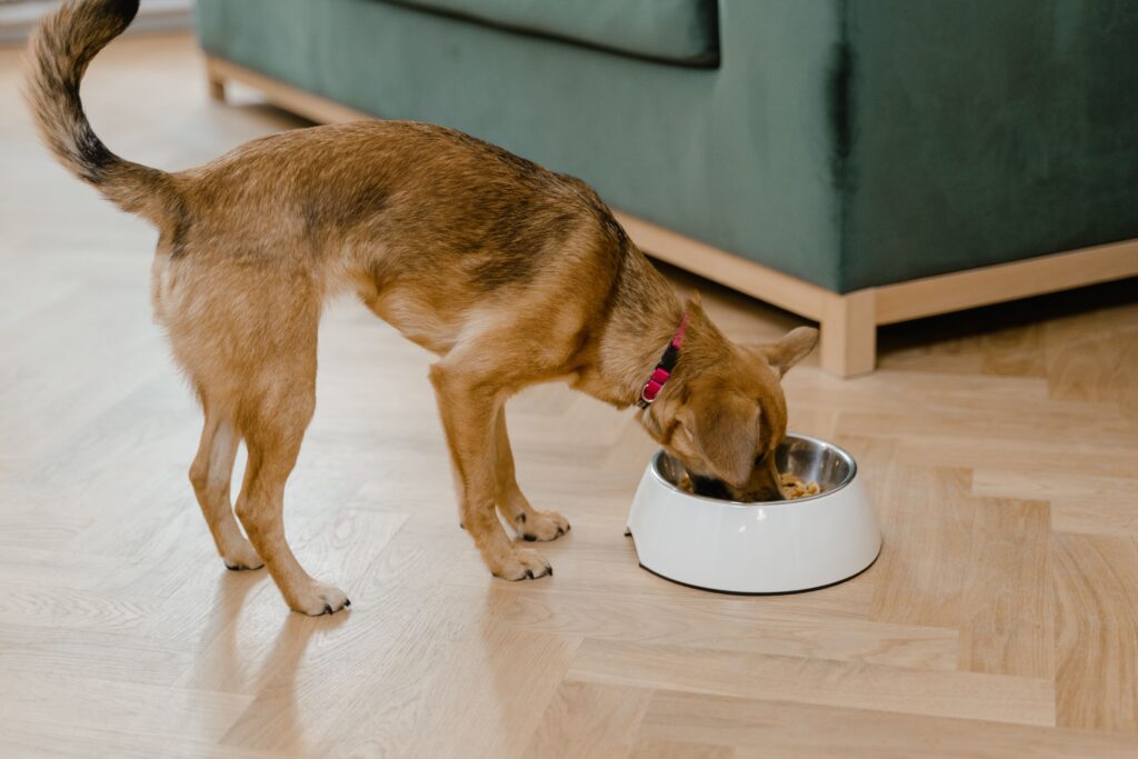
Increasing a dog’s water intake is essential to prevent kidney stones. A diet high in moisture content reduces kidney stone formation risk-switching to canned food or adding water to meals are practical strategies. Regular vet check-ups can help monitor kidney stone recurrence, with imaging recommended every 3–6 months for high-risk dogs so that new stones or more stones can be detected early, when they are still small enough for minimally invasive stone removal. These dogs should be monitored periodically with imaging and urinalysis to catch recurrence before clinical signs develop.
Managing Pain and Discomfort
Common signs of pain in dogs with kidney stones include flank sensitivity, abdominal pain, restlessness, appetite loss, vomiting, and lethargy. Dogs may show subtle behaviors such as reluctance to lie in certain positions, licking at their flank area, or generalized depression. Vomiting can indicate kidney stones in dogs, particularly when stones cause ureteral distension or renal capsule stretching.
Pain management typically involves non-steroidal anti-inflammatory drugs (NSAIDs) when renal function permits, supplemented by analgesics such as tramadol or gabapentin. For ureteral obstruction, pain tends to be more severe and may require more aggressive analgesia. Dose adjustments are essential in dogs with compromised kidney function, following guidelines from organizations like WSAVA and AAHA to protect remaining renal capacity.
Emergency Urinary Obstruction
Urinary obstruction from kidney stones is a critical emergency. Urinary blockages can lead to kidney damage through back-pressure (hydronephrosis), azotemia, dangerous hyperkalemia, and potentially fatal cardiac arrhythmias. This is especially dangerous in male dogs, whose narrower urethral anatomy increases obstruction risk.
Warning signs requiring immediate veterinary attention:
- Straining to urinate with little or no urine produced
- A distended, painful bladder on palpation
- Sudden onset of severe abdominal pain with vomiting and depression
- Complete inability to urinate
- Blood in the urine combined with systemic illness signs
Emergency intervention involves stabilizing the patient with intravenous fluids and correcting electrolyte imbalances, followed by imaging to locate the urinary blockage. Relief may involve catheterization, retrograde flushing, ureteral stent placement, or emergency surgical removal. As emphasized by PetMD’s emergency protocols, timely intervention is crucial to prevent permanent kidney damage-delay of even hours can mean the significant difference between recovery and irreversible renal loss.
Conclusion and Next Steps
Kidney stones in dogs range from incidental findings to urgent medical emergencies. The critical takeaways: calcium oxalate stones are the most common in dogs and require surgical intervention, struvite stones can be dissolved with diet and antibiotics when infection is the driver, and urate stones are linked to metabolic and liver conditions requiring long-term medical management. Regardless of stone type, increased water intake helps prevent kidney stones in dogs, and regular veterinary monitoring catches recurrence early.
Immediate steps for dog owners:
- Schedule a veterinary consultation if your dog shows any clinical signs-blood in urine, abdominal pain, changes in urination patterns, or lethargy
- Increase daily water intake through canned food, water additions to meals, or multiple fresh water stations
- Learn the warning signs of urinary obstruction and know your nearest emergency veterinary facility
- If your dog has had stones previously, discuss a preventive diet change and monitoring schedule with your veterinarian
For dogs with recurrent stones or breed-specific risk factors, consultation with a veterinary nephrology or urology specialist can provide advanced diagnostic testing and tailored prevention protocols. Related topics worth exploring include management of chronic kidney disease in dogs, nutritional strategies for urinary health, and breed-specific screening programs for genetic predisposition to urolithiasis.
Additional Resources
- Minnesota Urolith Center – Stone analysis services, epidemiological data on canine urinary stones, and breed-specific risk information
- ACVIM Consensus Recommendations on Urolithiasis in Dogs – Evidence-based treatment and prevention guidelines developed by the American College of Veterinary Internal Medicine (Lulich et al., 2016)
- Canadian Veterinary Urolith Centre research publications – Comprehensive analysis of 75,674 uroliths submitted between 1998–2014, including breed, sex, and mineral-type trends
- Prescription therapeutic diet resources – Consult your veterinarian about scientifically formulated diets designed for specific stone types; dietary management remains the cornerstone of long-term prevention in veterinary medicine