
Introduction
A skin biopsy is a diagnostic procedure in which a veterinarian removes a small tissue sample from a dog’s skin for microscopic examination by a veterinary pathologist. It is the most reliable method for reaching an accurate diagnosis when surface-level tests-cytology, skin scrapings, or bacterial culture-fail to identify the underlying cause of a skin condition.
This guide covers when skin biopsies are recommended, the different biopsy techniques used in veterinary medicine, what happens during and after the procedure, and how to interpret results. It is written for dog owners who have been told their pet may need a biopsy and want to understand every step of the process before, during, and after sample collection.
In short: A skin biopsy involves removing a small section of skin-including epidermis, dermis, and often subcutaneous tissue-so a pathologist can examine the cellular architecture under a microscope. Skin biopsies help differentiate between infections, cancers, and immune diseases, providing the definitive diagnosis that guides effective treatment.
After reading this guide, you will understand:
- When and why a veterinarian recommends skin biopsy for your dog
- The differences between punch biopsy, incisional elliptical biopsy, and excisional biopsy
- How to prepare your dog and what happens during the procedure
- What biopsy results mean and what treatment steps follow
- How to care for the biopsy site during recovery
Understanding Skin Biopsy Fundamentals
A skin biopsy is a diagnostic procedure for skin diseases in dogs. Unlike superficial tests such as skin scrapings or impression cytology, histopathology preserves the full structure of the tissue-the relationships between epidermal layers, the dermis, hair follicles, glands, and deeper structures. This architectural view allows a veterinary pathologist to identify patterns of disease that surface tests simply cannot reveal, from the acantholytic cells characteristic of pemphigus foliaceus to the mitotic figures that grade a mast cell tumor.
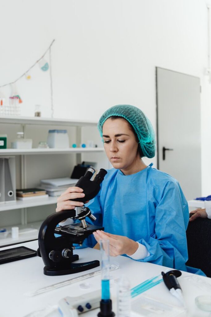
Biopsy samples are examined in a lab by a veterinary pathologist, who evaluates cellular detail, inflammation patterns, the presence of pathogens, and-in the case of tumors-margin status. This level of detail makes biopsy the gold standard when clinical signs point toward conditions that require tissue-level confirmation.
When Skin Biopsy is Recommended
Skin biopsies are performed on suspicious skin growths-nodules, masses, or lesions that appear suddenly, grow rapidly, or change in color or texture. Any suspicious skin growth that cannot be confidently identified through physical examination alone warrants biopsy to differentiate between benign and malignant tumors.
Equally important, skin biopsies are recommended for conditions that do not respond to treatment. Biopsy is indicated for lesions not responding to therapy after 3 weeks, including chronic dermatitis, persistent ulcerations, and recurrent pyoderma that fails to clear with appropriate antibiotics. Unusual or severe dermatosis may also indicate the need for a skin biopsy, particularly when clinical signs suggest immune mediated skin disease, deep fungal infection, or an atypical presentation.
These situations require tissue-level analysis because the differential diagnoses are broad-what looks like a simple infection on the surface may turn out to be a lymphoma, a drug reaction, or an autoimmune process that demands completely different therapy.
Types of Skin Conditions Diagnosed Through Biopsy
Skin biopsies can diagnose immune-mediated diseases such as pemphigus foliaceus, the most common autoimmune skin disease in dogs. Histopathology reveals characteristic subcorneal pustules with acantholytic cells that confirm the diagnosis. Other immune-mediated conditions-pemphigus vulgaris, discoid lupus erythematosus, bullous pemphigoid-each produce distinct histological patterns that a pathologist can identify.
Biopsies can identify infections, allergic diseases, and tumors. Deep bacterial infections, fungal organisms (dermatophytes, systemic mycoses), and mycobacterial infections that evade surface cytology are often diagnosed through tissue sections with special stains. Skin biopsies also diagnose conditions like cutaneous T-cell lymphoma, mast cell tumors, melanomas, and sarcomas, where histopathology determines tumor type, grade, and margin status.
Additionally, conditions like sebaceous adenitis, follicular dysplasia, and keratinization disorders are diagnosed through the specific architectural changes visible in biopsy samples-changes invisible to any surface-level test. Understanding which conditions biopsy can reveal makes it clear why technique selection matters.
Biopsy Techniques and Methods
The technique a veterinarian chooses depends on lesion size, location, the suspected diagnosis, and whether complete removal is both possible and desirable. Common biopsy techniques include punch biopsies and excisional biopsies, with incisional methods filling an important middle ground.
Punch Biopsy
A skin punch biopsy uses a cylindrical cutting instrument-essentially a small, circular scalpel blade-to obtain a full-thickness core of skin. The punch is pressed against the skin and rotated in one direction until it cuts through the epidermis, dermis, and into the subcutaneous tissue, yielding a uniform columnar sample.
Punch biopsies typically use a 6-8 mm punch size. While smaller punches (2-4 mm) exist, a 6-8mm punch provides optimal biopsy size with enough tissue for the pathologist to evaluate architectural patterns without sampling error. Most skin biopsies performed for inflammatory dermatoses, alopecic lesions, and suspected autoimmune diseases use this technique because it is fast, minimally invasive, and commonly performed under local anesthesia as an outpatient procedure.
Incisional Biopsy
An incisional elliptical biopsy removes a wedge-shaped or elliptical portion of a larger lesion using a scalpel blade and iris scissors. This technique captures deeper tissue than a punch can reach and preserves the transition zone between affected and normal skin-critical for diseases where the junction reveals the most diagnostic information.
Incisional biopsies are preferred over punch biopsy when lesions are too large for complete removal, when disease extends deep into the subcutaneous tissue or panniculus, or when a wedge biopsy capturing the lesion border provides superior architectural information. The trade-off is a larger wound requiring suture closure and, typically, sedation or general anesthesia.
Excisional Biopsy
An excisional biopsy removes the entire lesion with surrounding margins of normal tissue. This approach is both diagnostic and potentially therapeutic-if the pathologist confirms clean margins, the procedure may constitute a cure for benign tumors or early-stage malignancies.
Excisional biopsy is best suited for smaller, well-circumscribed masses where complete removal is feasible without functional or cosmetic compromise. The technique requires careful surgical planning, including consideration of closure methods (primary closure, skin flaps, or grafts for larger defects), and generally demands general anesthesia.
Choosing between these techniques ultimately depends on what the veterinarian suspects, the lesion’s characteristics, and the information needed. With technique selected, the focus shifts to execution.
Practical Implementation and Procedure
Understanding what happens during a veterinary visit for skin biopsy helps dog owners prepare their pet and set realistic expectations for the day.
Pre-Biopsy Preparation Steps
Proper preparation directly influences the quality of biopsy samples and the likelihood of an accurate diagnosis. Active disease is required for an accurate biopsy diagnosis-sampling old, scarred, or end-stage lesions often yields nondiagnostic results.
- Discontinue interfering medications: Corticosteroids should be suspended 2-8 weeks before biopsy, as they suppress the immune response and can mask the histological features a pathologist needs to see. Systemic antibiotics and antifungals should also be paused when safely possible-discuss timing with your veterinarian.
- Select primary lesions during active disease: The veterinarian will identify the most representative lesions-fresh crusts, intact pustules, or active margins of expanding lesions rather than areas of chronic scarring. Three to five biopsy sites are recommended for accurate diagnosis, ideally capturing different lesion morphologies.
- Determine anesthesia requirements: The procedure may use local anesthesia and can be performed as an outpatient procedure for punch biopsies. A local anesthetic is commonly used during skin biopsies, often lidocaine buffered with sodium bicarbonate to reduce injection discomfort. Larger incisional or excisional biopsies on sensitive areas, or in patients with high anxiety, may require sedation or general anesthesia.
- Prepare the biopsy site carefully: Avoid using electric clippers to prepare biopsy sites, as aggressive clipping can remove diagnostic crusts or disrupt fragile epidermal structures. Instead, gently clip the hair with fine scissors, preserving the lesion surface. Avoid aggressive antiseptic scrubbing for the same reason-clean the area gently without destroying surface pathology.
Technique Comparison
Criterion | Punch Biopsy | Incisional Biopsy | Excisional Biopsy |
|---|---|---|---|
Sample size | Small section (6-8 mm core) | Moderate (elliptical or wedge) | Entire lesion with margins |
Depth | Full-thickness to superficial subcutis | Deep, including subcutaneous tissue | Complete, all layers |
Anesthesia | Local anesthetic typically sufficient | Sedation or general anesthesia | General anesthesia usually required |
Closure | Often 1-2 sutures or heals by secondary intention | Suture required | Suture, flaps, or grafts may be needed |
Best for | Inflammatory skin diseases, alopecia, small lesions | Large or deep lesions, transition zones | Small masses, suspected neoplasms |
Therapeutic potential | Diagnostic only | Diagnostic only | Diagnostic and potentially curative |
Typical cost range (U.S.) | $300–$900 | $500–$1,200 | $1,000–$2,500+ |
The right choice balances diagnostic needs against patient welfare, lesion characteristics, and the sensitivity of the anatomic site.
Sample Processing and Submission
After tissue is obtained, proper handling preserves the diagnostic value of every sample:
- Submit biopsy samples in 10% formalin for fixation, using a formalin volume at least ten times the volume of the tissue. Delays or inadequate fixation degrade cellular architecture and can render samples nondiagnostic.
- Handle tissue gently to avoid crush artifact-grasp specimens by the fat at the base rather than the epidermis. Excess blood should be blotted carefully, but do not compress the tissue with forceps.
- Provide complete clinical history with biopsy samples, including lesion duration, distribution, prior medications, differential diagnoses, and response to previous therapy. Detailed clinical history significantly increases a pathologist’s ability to reach a definitive diagnosis.
- Notify the pathologist of any additional crusts submitted separately, as these may contain diagnostic material (acantholytic cells, fungal elements, bacterial colonies).
- If special testing is required-direct immunofluorescence, bacterial culture, or fungal culture-send separate, unfixed portions in appropriate transport media. Formalin kills organisms and destroys antigens needed for immunofluorescence.
With samples properly submitted, the waiting period begins-but understanding what comes back in the report matters just as much as what went into the jar.
Common Challenges and Solutions
Several practical issues can compromise biopsy quality or complicate the diagnostic process. Knowing them in advance helps both veterinarians and owners achieve better outcomes.
Inadequate Sample Quality
Crushing artifacts, thermal damage from electrocautery, and undersized samples are the most common reasons biopsies fail to yield a diagnosis. The solution is disciplined technique: use a 6-8mm punch for optimal tissue volume, rotate the punch in one direction to maintain a clean cut, handle tissue exclusively by the subcutaneous fat, and avoid cautery within the sample zone. If a small amount of hemorrhage occurs, apply gentle pressure rather than cauterizing the specimen.
Multiple Lesion Types Present
Dogs often present with several morphologically distinct lesions-nodules alongside crusts, alopecic lesions next to pigmented plaques. Collect 3-5 biopsies from different locations for accuracy, labeling each sample with its anatomic site, lesion type, and whether it represents early, active, or chronic disease. This strategic sampling gives the pathologist the full picture rather than a fragment.
Patient Anxiety or Difficult Locations
Biopsies on the face, paw pads, or near joints present both access challenges and heightened patient sensitivity. Appropriate sedation protocols-ranging from light oral sedation to injectable combinations-paired with local anesthetic nerve blocks can address both movement and pain. For particularly anxious patients or complex excisional procedures, general anesthesia ensures both safety and sample quality. AI-powered symptom checkers can help owners assess their dog’s condition before the veterinary visit, reducing uncertainty about whether the clinical signs warrant biopsy.
Interpreting Results and Next Steps
Skin biopsy results typically take one to two weeks to process, though complex cases requiring special stains (PAS for fungi, Ziehl-Neelsen for mycobacteria) or immunohistochemistry may take longer. The histopathology report will include a morphological description, a diagnosis or ranked differential diagnoses, and-for tumors-information about cell type, grade, mitotic index, and margin status.
Results directly guide treatment decisions:
- Neoplastic diagnoses determine whether surgical follow-up, chemotherapy, or radiation therapy is needed. Clean excisional margins may mean no further treatment is required; incomplete margins often prompt re-excision or adjuvant therapy.
- Immune mediated skin disease confirmed by biopsy typically requires systemic immunosuppressive therapy-often starting with corticosteroids and potentially adding steroid-sparing agents for long-term management.
- Deep infections identified through biopsy and concurrent culture guide targeted antimicrobial therapy rather than empirical broad-spectrum treatment, supporting antimicrobial stewardship in veterinary dermatology.
- Inconclusive results occur when samples capture only chronic, nonspecific changes. In these cases, repeat biopsies from newly active primary lesions, additional testing such as immunofluorescence or PCR, or referral to a veterinary dermatology specialist may be necessary.
Post-procedure monitoring includes daily inspection of the biopsy site for signs of infection (redness, swelling, discharge), keeping sutures clean and dry, preventing licking with an E-collar, and restricting activity for 7-14 days depending on wound size. Suture removal is typically performed at 10-14 days. For owners tracking their dog’s recovery or wanting to better understand diagnostic results, AI-assisted tools can help contextualize findings alongside blood work and other test results.
Advances in artificial intelligence in veterinary medicine are also beginning to influence dermatopathology, with digital pathology platforms and AI tools under development to assist pathologists in detecting artifacts and improving diagnostic consistency across laboratories.
Frequently Asked Questions
How much does a skin biopsy cost for a dog? In the United States, the typical cost ranges from $300 to $1,200 for most skin biopsies, with many clustering near $700. Punch biopsies fall in the lower range ($300–$900), while surgical excisional biopsies can reach $1,000–$2,500 or more depending on complexity, location, and whether general anesthesia is required. Pet insurance policies that cover diagnostics may reimburse a portion of these costs.
Is a skin biopsy painful for my dog? A local anesthetic is commonly used during skin biopsies, numbing the area so the dog feels pressure but not sharp pain. For larger procedures or anxious patients, sedation or general anesthesia ensures comfort throughout. Post-procedure pain is typically mild and managed with prescribed analgesics or NSAIDs for a few days.
How long does healing take after a skin biopsy? Small punch biopsy sites often heal within 7-10 days. Sutured incisional or excisional sites generally require 10-14 days before suture removal. Activity restriction-limiting running, swimming, and rough play-is recommended during the healing period to prevent wound dehiscence. Complications such as infection are rare, particularly for punch and incisional biopsies.
What happens if biopsy results are inconclusive? Some diseases, particularly allergic conditions or early-stage endocrine disorders, produce nonspecific histological changes. The veterinarian may recommend repeat biopsies from different or newer lesions, additional diagnostics (immunofluorescence, PCR, culture), or a second opinion from a board-certified dermatopathologist. Three to four biopsy sites should be sampled for accurate results, reducing the risk of sampling error from the start.
When should I seek emergency care after a biopsy? Contact your veterinarian promptly if the biopsy site shows significant bleeding that doesn’t stop with gentle pressure, increasing redness or swelling beyond the first 24 hours, foul-smelling discharge, or if your dog develops fever, lethargy, or refuses to eat. While serious complications are uncommon, early intervention prevents escalation.