Neurological symptoms in dogs send thousands of pet owners to veterinary specialists each year, with many of these cases requiring magnetic resonance imaging to confirm a diagnosis. Dog MRI images reveal what X-rays and physical exams cannot—the intricate details of brain tissue, spinal cord integrity, and soft tissue abnormalities that dictate treatment paths and prognosis.
Yet the journey from scan to actionable insight involves multiple steps: image acquisition, radiologist interpretation, report generation, communication with referring veterinarians, and integration into the patient’s broader health narrative.
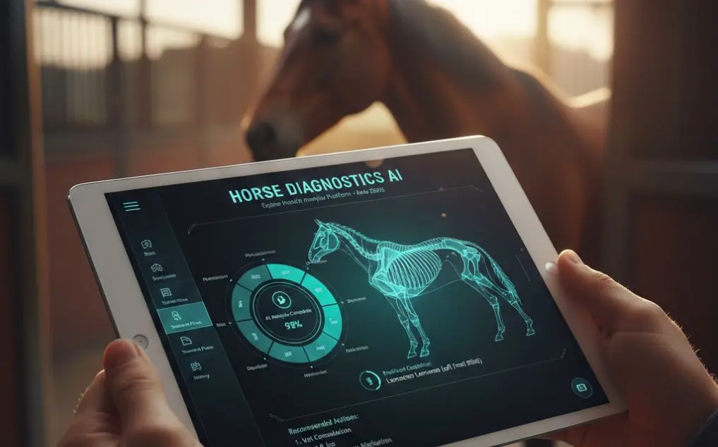
This is where artificial intelligence transforms veterinary diagnostics. CompanAIn’s multi-agent AI platform processes MRI findings alongside lab work, clinical notes, and historical data to deliver comprehensive, context-aware insights that help veterinarians make faster, more informed decisions. By organizing complex diagnostic information into digestible timelines and flagging emerging patterns, AI bridges the gap between raw imaging data and proactive patient care.
Why Dog MRI Images Matter
Magnetic resonance imaging stands apart from other diagnostic modalities because it excels at visualizing soft tissues—the very structures most vulnerable to neurological disease. When a dog presents with sudden paralysis, seizures, balance loss, or unexplained pain, MRI often provides the definitive answer.
Common conditions requiring MRI evaluation include intervertebral disc disease (IVDD), particularly prevalent in long-bodied breeds like Dachshunds and Corgis. Research demonstrates that MRI achieves sensitivity exceeding 98% for detecting IVDD, significantly outperforming other imaging methods.
Brain tumors, another frequent indication, appear clearly on MRI sequences that distinguish between gray matter, white matter, and abnormal masses. Spinal cord compression from herniated discs, tumors, or inflammation becomes visible with detail impossible through conventional imaging.
Stroke events in dogs, though less common than in humans, require MRI confirmation to differentiate from other acute neurological episodes. Inflammatory myelopathy—conditions where the immune system attacks nervous tissue—shows characteristic patterns on MRI that guide treatment with immunosuppressive therapy rather than surgery.
The advantage over computed tomography (CT) or radiography lies in MRI’s superior soft tissue contrast. While CT excels at bone detail and offers faster acquisition, MRI reveals subtle edema, inflammation, and early degenerative changes that CT might miss entirely.
For neurological cases, this distinction often determines whether a patient receives appropriate treatment in time to prevent permanent damage. Dogs with IVDD, for instance, experience drastically different outcomes based on intervention speed.
Studies show that dogs with intact pain sensation have approximately 80% recovery rates following surgical intervention. However, timing remains critical—delays reduce favorable outcomes significantly, making rapid, accurate MRI interpretation essential for optimal patient care.
How AI Enhances MRI Image Analysis
Traditional MRI interpretation follows a sequential workflow: the imaging facility generates sequences, a radiologist examines hundreds of slices, identifies abnormalities, measures lesions, compares findings to previous studies, and produces a detailed report.
This process, while thorough, introduces variables—time constraints, fatigue-related oversights, and the inherent challenge of pattern recognition across massive datasets.
CompanAIn’s multi-agent AI system approaches this differently. Rather than replacing the radiologist’s expertise, it augments the entire diagnostic pipeline by processing MRI reports, measurements, and findings the moment they’re available.
The platform’s Specialized Pathologist Agent analyzes imaging results within the context of the patient’s complete medical history—previous labs, medication responses, symptom progression, and breed-specific risk factors.
This contextual analysis provides something traditional interpretation workflows often lack: longitudinal pattern recognition. An MRI might show mild spinal cord compression that, in isolation, suggests conservative management.
But when AI cross-references this finding with subtle changes in recent bloodwork, declining mobility noted in clinical records, and progression patterns from similar cases, it may flag the need for more aggressive intervention.
Research examining AI assistance in veterinary imaging has demonstrated measurable reductions in diagnostic error rates, primarily by catching subtle findings that human eyes might dismiss as artifacts or normal variation.
Consistency represents another advantage—AI doesn’t experience the fatigue or cognitive load that affects human performance during marathon reading sessions.
For veterinary practices without in-house radiology specialists, AI analysis offers access to expertise that might otherwise require costly referrals or multi-day delays waiting for teleradiology reports.
CompanAIn’s system processes findings and generates preliminary assessments that help general practitioners determine urgency, guide client conversations, and coordinate specialist consultations more effectively.
The platform’s integration capabilities mean MRI findings don’t exist as isolated PDFs buried in email attachments. Instead, they populate the patient’s living health timeline, positioned chronologically alongside relevant lab values, medication adjustments, and symptom changes.
This unified view transforms how veterinarians approach complex cases, enabling them to spot connections that fragmented records obscure.
Licensed veterinarians review all critical alerts and low-confidence assessments generated by the AI, ensuring that flagged concerns receive appropriate clinical judgment before reaching the primary care team. This human-AI collaboration model preserves professional oversight while leveraging computational advantages in data processing and pattern recognition.
CompanAIn's Platform Features
The architecture behind CompanAIn’s diagnostic support system relies on specialized AI agents working collaboratively rather than a single monolithic model attempting to handle every task.
The Data Aggregator agent imports and standardizes information from multiple sources—radiology reports, laboratory systems, clinical notes, and owner-submitted documents. This normalization process ensures consistency regardless of source format.
The Health Analyzer agent examines these aggregated records for trends, correlations, and anomalies. When new MRI findings arrive, this agent evaluates them against the patient’s baseline, identifies deviations from expected values, and calculates risk scores for various differential diagnoses.
The Recommendation Engine synthesizes insights from other agents to generate actionable care plans. Following an MRI revealing early disc degeneration, for example, it might suggest weight management protocols, exercise modifications, and monitoring schedules tailored to the individual dog’s lifestyle and breed predispositions.
Security and compliance underpin every function. End-to-end encryption protects patient data during transmission and storage. The platform adheres to industry-leading data protection standards, ensuring that sensitive medical information remains confidential and accessible only to authorized parties.
The user interface prioritizes clarity over complexity. Veterinarians navigate intuitive dashboards displaying color-coded alerts, interactive timelines, and detailed reports without requiring extensive training.
Non-radiologist practitioners can quickly grasp key findings, understand their clinical significance, and communicate results to pet owners with confidence.
Report generation happens automatically, translating technical imaging findings into formats appropriate for different audiences. Referring veterinarians receive comprehensive clinical summaries with treatment recommendations. Pet owners access simplified explanations highlighting what the findings mean for their dog’s health and outlining next steps.
Clinical Applications and Case Examples
Emergency scenarios demonstrate AI-assisted imaging triage at its most valuable: a seven-year-old German Shepherd arrives at an emergency clinic with acute paralysis. MRI reveals severe spinal cord compression at L2-L3.
CompanAIn’s system processes the radiologist’s preliminary findings within minutes, flags the case as requiring immediate surgical consultation based on compression severity and symptom duration, and generates a surgical planning report highlighting anatomical landmarks and decompression targets.
The speed advantage—compared to waiting hours for formal radiology reports—can mean the difference between recovery and permanent disability.
Surgical planning benefits from AI’s ability to synthesize imaging data with surgical outcomes databases. When an MRI shows a brain mass, the platform references similar cases involving the same breed, tumor location, and size parameters.
It presents success rates for different surgical approaches, radiation protocols, and palliative care options, giving veterinary surgeons evidence-based context for treatment discussions.
Post-operative monitoring leverages sequential imaging analysis. After a dog undergoes surgery for IVDD, follow-up MRI at six weeks might show subtle inflammation around surgical hardware.
CompanAIn’s comparison tools highlight changes from the immediate post-operative scan, flag unusual healing patterns, and suggest whether findings represent normal recovery or developing complications.
Diagnostic differentiation—distinguishing between conditions with overlapping presentations—becomes more reliable when AI considers the complete clinical picture. An MRI showing brain inflammation could indicate infectious meningitis, immune-mediated encephalitis, or early-stage lymphoma.
By analyzing concurrent lab abnormalities, response to empirical treatments, and progression timing, AI can weigh differential diagnoses more accurately than imaging findings alone allow.
Referral optimization occurs when general practitioners use AI analysis to determine which cases require specialist intervention versus those manageable with primary care.
An MRI showing mild age-related brain changes might not warrant neurology referral if AI analysis confirms findings consistent with normal aging and unrelated to presenting symptoms. Conversely, subtle findings that AI flags as concerning despite initially appearing benign can prompt timely specialist engagement.
Future of Veterinary Diagnostic AI
The trajectory of AI in veterinary medicine points toward increasingly sophisticated integration across diagnostic modalities. Current systems analyze imaging, laboratory, and clinical data separately.
Next-generation platforms will synthesize real-time information from multiple sources simultaneously—correlating MRI findings with bloodwork trends, genetic predisposition data, and environmental factors to generate truly holistic health assessments.
Predictive analytics represent the next frontier. Rather than reacting to abnormalities after they appear on imaging, AI will identify risk patterns suggesting disease before structural changes become visible.
Early markers in metabolic panels, subtle gait changes noted in activity data, and genetic risk factors could trigger preventive imaging protocols, catching conditions like degenerative myelopathy or brain tumors in treatable stages.
Monitoring subtle neurological changes can also help identify early signs of musculoskeletal conditions like dog arthritis and age-related changes, offering timely intervention for long-term joint health.
Accessibility improvements may prove most impactful. Rural veterinary practices and underserved regions often lack access to advanced imaging and specialist interpretation.
Cloud-based AI platforms democratize expertise, enabling a small-town clinic to provide diagnostic quality previously available only at university teaching hospitals. This geographic leveling matters particularly for conditions where treatment timing determines outcomes.
Integration with wearable technology and home monitoring systems will close the loop between clinical diagnostics and daily life. MRI might reveal early arthritis, prompting the AI to monitor activity patterns, sleep quality, and movement symmetry through connected devices.
Changes in these metrics could trigger alerts for follow-up imaging, creating a continuous care model that catches progression before symptoms become severe.
The evolution from reactive to proactive care—CompanAIn’s core mission—accelerates as AI capabilities expand. Today’s systems excel at organizing information and highlighting patterns. Tomorrow’s will predict disease trajectories, recommend preventive interventions, and personalize treatment protocols with precision impossible through traditional approaches.
Transform Your Practice with AI-Powered Diagnostics
Dog MRI images provide unparalleled insight into neurological health, but their value depends on rapid, accurate interpretation integrated into comprehensive patient care.
CompanAIn’s multi-agent AI platform bridges the gap between imaging data and actionable clinical decisions, helping veterinarians deliver faster diagnoses, evidence-based treatment plans, and proactive health management.
Whether you’re a specialist seeking enhanced workflow efficiency, a general practitioner navigating complex neurological cases, or a pet owner researching diagnostic options for your dog, AI-powered analysis represents the future of veterinary medicine.
CompanAIn makes advanced diagnostic support accessible, affordable, and seamlessly integrated into existing workflows.
Discover how veterinary practices are improving diagnostic accuracy and patient outcomes with CompanAIn’s AI platform.
Frequently Asked Questions
How accurate is AI analysis compared to board-certified veterinary radiologists?
CompanAIn’s AI platform demonstrates high accuracy for common pathologies including IVDD, brain masses, and inflammatory conditions. The platform functions as a collaborative tool rather than radiologist replacement, providing rapid preliminary assessment and second-opinion capabilities.
Licensed veterinarians review all critical findings, combining AI’s pattern recognition strengths with human clinical judgment for optimal diagnostic reliability.
What breeds are most prone to conditions requiring MRI?
Dachshunds, Corgis, and other chondrodystrophic breeds face elevated IVDD risk due to body conformation. Large breeds including German Shepherds and Labrador Retrievers show predisposition to degenerative myelopathy and spinal tumors.
Brachycephalic breeds like French Bulldogs and Pugs experience higher rates of brain abnormalities related to skull shape. Golden Retrievers demonstrate increased brain tumor incidence.
Breed-specific risk factors inform CompanAIn’s analysis algorithms, providing context for interpreting findings.
How much does a dog MRI cost with AI analysis?
Standard canine MRI procedures range from $1,500 to $3,500 depending on body region, sedation requirements, and geographic location.
CompanAIn’s AI analysis adds value by reducing diagnostic timelines, minimizing repeat imaging needs, and enabling more targeted treatment planning. The efficiency gains often offset costs through fewer complications, shorter hospital stays, and improved outcomes that prevent expensive secondary interventions.
Can pet owners access their dog's MRI images?
Yes. CompanAIn provides detailed, owner-friendly reports summarizing MRI findings in accessible language. Pet owners receive visual aids, annotated images highlighting key areas, and clear explanations of what results mean for their dog’s health.
The platform facilitates image sharing for second opinions or specialist referrals, ensuring owners remain informed partners in their pet’s care journey.
How long does AI analysis take?
Initial processing typically completes within 24-48 hours of receiving imaging reports. Urgent cases flagged by preliminary assessment receive expedited review.
The platform’s real-time integration means insights become available as soon as radiologist reports enter the system, dramatically faster than traditional workflows requiring manual record assembly and review.
Is the MRI procedure safe for dogs?
MRI uses magnetic fields and radio waves rather than ionizing radiation, making it non-invasive and safe for repeated use. Dogs require sedation or general anesthesia to remain motionless during image acquisition, which carries standard anesthetic risks managed through pre-procedure health screening and continuous monitoring.
The diagnostic benefits typically far outweigh these minimal risks, particularly for conditions where accurate imaging determines treatment success.