
Your dog’s skin tells a story. Redness, rashes, hair loss, crusty ears, or a mysterious new lump can signal anything from a mild allergy to a condition that needs urgent veterinary attention. The challenge is knowing which is which-especially when fur hides the evidence. AI image analysis for dermatological issues in dogs is changing how pet owners spot problems early and how veterinarians manage skin cases, turning a smartphone photo into actionable health guidance.
Fast answer: how CompanAIn can help with your dog's skin problem today
CompanAIn uses artificial intelligence and image analysis to flag common dog skin diseases from photos you take at home. AI dermatology tools analyze images for skin conditions in seconds, giving you rapid preliminary insights into what might be going on before your next vet appointment.
Here are concrete examples of skin concerns you can check right now:
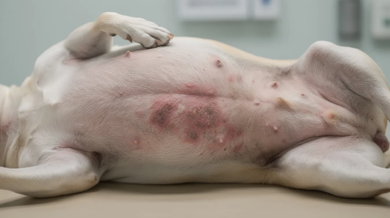
- A red rash on your dog’s belly or groin
- Hair loss around the tail base, flanks, or face
- A moist, oozing hot spot on the neck or thigh
- A new lump or bump that appeared over the past few weeks
- Crusty or scabby edges on the ear flaps
- Black spots or darkened skin in hairless areas
- A circular bald patch that looks like suspected ringworm
When you upload a clear photo, the AI analyzes the image-evaluating fur coverage, color, texture, and visible skin lesions-and suggests likely skin conditions. It then assigns an urgency rating: watch at home, see your vet within 24–48 hours, or seek same-day emergency care. AI offers rapid preliminary insights into canine skin conditions, helping you decide how quickly to act.
This is educational guidance, not a formal veterinary diagnosis. AI can flag common skin conditions in pets and provide personalized health insights for pet owners, but all serious signs-pain, fever, rapid worsening-still require an in-person vet visit. When in doubt, always call your veterinarian.
Why dog skin diseases are so common (and easy to miss at home)
Dermatology consistently ranks among the top three reasons dog owners visit veterinarians across Europe and North America. Practice data from large vet groups in 2023 confirmed that skin allergies topped the list of canine insurance claims, with ear infections coming in a close second. In a UK study of veterinary advice networks, 81% of dermatology-related requests involved canine cases, with itchy skin, alopecia, and crusting leading the way.
There are over 200 recognized skin diseases in dogs, ranging from mild flea allergy to serious skin cancer, making visual diagnosis and treatment planning challenging even for experienced clinicians. Many canine skin diseases present similar symptoms visually, complicating diagnosis for everyone involved. Skin irritation is the most common reason for rashes, but the underlying cause can vary wildly.
Here’s why problems are easy to miss at home:
- Fur hides early lesions. A developing rash under a thick double coat can go unnoticed for weeks.
- Early rashes look similar. Mild redness from allergies, infections, and parasites can appear nearly identical in the beginning.
- Gradual hair loss seems like “just shedding.” A golden retriever with year-round paw licking that owners think is a behavioral quirk, not an allergy, is a classic example.
- Seasonal patterns confuse people. Flares that come and go with spring pollen look like the problem “resolved itself.”
- Dogs can’t tell you they itch. Subtle behaviors like face rubbing, belly licking, or ear scratching are easily dismissed.
Chronic atopic dermatitis and recurrent ear infections aren’t cosmetic inconveniences-they are major quality-of-life issues that affect sleep, appetite, and temperament in dogs.
What AI dermatology means for dogs
AI dermatology, in the context of companion animals, refers to using artificial intelligence and machine learning to interpret photos of the skin and associated areas-ears, paws, nails-to recognize patterns of skin conditions. AI image analysis identifies dog skin problems using computer vision models trained on thousands of annotated veterinary images. These tools aid in diagnosing skin diseases in dogs and cats by matching visual patterns to known dermatological conditions.
Modern deep learning models, particularly convolutional neural networks, are trained on tens of thousands of annotated dog skin images-similar in principle to the human AI dermatology tools now used for skin cancer screening. CompanAIn focuses specifically on companion animals and integrates image analysis with the dog’s health history and timeline, including itch patterns, diet changes, and previous lab results, for richer context. AI assists veterinarians by acting as a secondary diagnostic support system, not a replacement.
Here’s how to think about the scope:
- AI provides probabilities, confidence scores, and new insights-it narrows the list of possible conditions.
- Veterinarians integrate the exam, lab tests, history, and clinical judgment to reach a formal diagnosis.
- AI tools can flag common skin conditions in pets and improve the accuracy of veterinary diagnostics, but they cannot perform skin scrapings, cultures, or biopsies.
- The AI-powered pet symptom checker complements image analysis by correlating photos with reported symptoms for a fuller picture.
How AI skin image analysis works step by step
Here’s what happens between uploading a photo and getting results in the CompanAIn app. Image capture utilizes digital photos for analysis of skin conditions, and the entire process is designed to be simple for pet owners.
- Select the body area and concern. You choose the affected area (belly, paw, ear, flank) and briefly describe your concern-itch, lump, rash, or hair loss.
- Upload a photo. Snap or upload a JPG or PNG image. The app checks basic quality (focus, lighting, resolution) before proceeding.
- AI processes the image. The model segments the relevant skin area, analyzing fur coverage, color distribution, texture, and visible lesions. It can distinguish redness, papules, pustules, scaling, crusting, ulceration, and areas of hair loss, even through partial fur coverage.
- Pattern matching against labeled data. The algorithm compares extracted features against labeled veterinary data-thousands of confirmed cases annotated by veterinary professionals.
- Contextual refinement. CompanAIn combines the photo with optional questionnaire data: itch level on a severity index, duration of the problem, recent diet or flea product changes, and the dog’s age, breed, and history.
- Structured report generation. You receive a report listing likely conditions with confidence scores, visual highlights on the image showing the affected area, and a clear action recommendation (watch, schedule vet visit, or urgent care).
AI enables faster and more consistent image assessments than manual review alone, giving you a starting point while your vet appointment is still days away.
Taking clear photos of your dog's skin for AI analysis
Image quality significantly affects the accuracy of AI diagnostic tools. A blurry, dark, or poorly framed photo can shift which skin conditions are suggested and reduce confidence in the results.
Follow these steps for the best results:
- Use good natural daylight or soft indoor light. Avoid direct flash, which creates harsh reflections-especially on dark coats.
- Hold your phone 10–20 cm from the skin surface. The lesion should be centered and in sharp focus.
- Part the fur gently to expose the skin on areas like the belly, tail base, armpits, and paws. If your vet has previously approved it, you can trim long fur around (not over) the lesion for a clearer view.
- Take multiple angles if the lesion is large or irregularly shaped.
For tricky areas, adjust your approach:
- Ears: Photograph both the inside and outside of the ear flap (pinna). Gently fold the ear back and use diffused light to avoid shadows in the ear canal.
- Paw pads: Lift the paw and photograph from below. Capture the spaces between toes separately.
- Nose and around eyes: Move slowly, keep the dog calm, and use a helper to steady the head. Prioritize safety and comfort over the perfect shot.
For example, if you spot a red, moist patch on your Labrador’s thigh in July, photograph it in shade rather than direct sunlight. Place a coin beside the lesion for scale-this helps track growth over time.
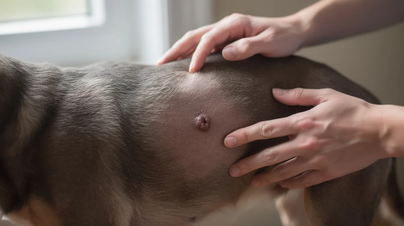
Which dog skin problems AI can help assess from photos
Visual AI works best for diseases where the skin appearance is distinctive and visible on the surface. AI can analyze skin images for various conditions, and AI can classify skin lesions with over 80% accuracy for the most common presentations. Here are major categories the tool can flag patterns for:
- Allergic rashes: Diffuse redness on the belly, groin, paws, and ear flaps, often with itch-related excoriation. Skin irritation from environmental or food allergens is among the most frequent findings.
- Hot spots (acute moist dermatitis): Sharply defined, wet, oozing patches that appear suddenly, usually on the neck, rump, or thighs.
- Ringworm: Circular areas of hair loss with scaly or crusty borders, sometimes with a ring-shaped pattern.
- Mange: Crusty, thickened skin on ear margins and elbows (sarcoptic) or patchy facial hair loss (demodectic).
- Flea allergy dermatitis: Hair loss and redness concentrated on the tail base, lower back, and hind legs, often with visible flea dirt.
- Bacterial skin infections (pyoderma): Red bumps, pustules, collarettes, and crusts-sometimes mistaken for “just a rash.”
- Yeast dermatitis: Greasy, reddish-brown staining between toes or in skin folds, with a mushy or waxy texture.
- Signs suggestive of endocrine disease: Patterned, symmetric hair loss on flanks or trunk, skin darkening, and poor coat regrowth-subtle clues that AI can flag for further vet work-up.
CompanAIn’s dataset focuses on common canine presentations collected between 2018 and 2026, with ongoing updates as new cases are labeled by veterinary professionals. Some internal diseases only show subtle external signs, so the AI may recommend further vet work-up even when the rash looks mild.
Hair loss in dogs: how AI distinguishes patterns
“My dog has hair loss” is one of the most frequent queries from pet owners, and the causes span allergies, parasites, hormonal disease, and behavioral patterns. AI helps by recognizing distinct visual signatures:
- Localized circular hair loss with scaly borders → possible ringworm or demodectic mange.
- Patchy hair loss with crusts and scabs → may indicate sarcoptic mange, bacterial infection, or trauma.
- Symmetric flank or tail-base hair loss without intense itch → raises suspicion of endocrine disease (hypothyroidism, Cushing’s) or flea allergy.
- Over-groomed areas from itch or pain → often allergic or behavioral, with broken stubble rather than smooth bald skin.
Combining image features-lesion edges, color, scaling, presence of pustules-with the owner-reported itch level helps the algorithm separate endocrine hair loss (typically low itch) from intense allergic or parasitic skin disease. CompanAIn’s report highlights red-flag patterns such as rapidly expanding bald areas, ulceration, or signs of systemic illness that should trigger direct vet consultation.
Allergic skin disease and atopic dermatitis in dogs
Atopic dermatitis is a chronic, inflammatory skin disease that often starts before age three, with strong links to lifelong itch, recurrent ear infections, and secondary bacterial or yeast infections. It is one of the most common chronic skin conditions in dogs.
Typical atopic dermatitis patterns AI can flag include:
- Red, inflamed paws with brown saliva staining from constant licking
- Recurrent ear redness and waxy discharge
- Belly and groin rashes that flare and partially resolve
- Hair loss on the muzzle and around the eyes
- Thickened, darkened skin that develops over months to years (lichenification)
Food allergy and environmental atopy can overlap visually, though food allergy tends to be non-seasonal while environmental atopy often flares in spring and autumn. AI can’t definitively separate them, but it can suggest patterns to discuss with your vet-a conversation that may lead to a dietary elimination trial or allergy testing.
Breeds that appear frequently in dermatology datasets include French Bulldogs, West Highland White Terriers, Labradors, and Golden Retrievers, though any dog can be affected regardless of breed or age. CompanAIn can track flare frequency and response to treatment in the health timeline, giving new insights into triggers across months and seasons.
Hot spots and acute moist dermatitis
Hot spots are sudden, intensely itchy, moist, oozing skin lesions that can double in size within hours. They’re especially common in thick-coated dogs during warm, humid weather-think a Bernese Mountain Dog in August.
On images, hot spots show distinctive features:
- A sharply demarcated red or raw patch with a wet, glistening surface
- Loss of hair directly over the lesion, with surrounding fur matted or stained
- Edges that look “punched out” compared to the gradual borders of many allergic reactions
- Common locations: neck, rump, and outer thighs
AI can differentiate hot spots from ringworm or mild skin irritation by analyzing texture, border sharpness, and the moisture shine visible in the photo. The urgency guidance in CompanAIn’s report typically recommends same-day vet assessment for large or rapidly growing lesions, or if the dog is in obvious distress-because hot spots can become painful, infected, and much harder to treat if left even 24 hours.
Parasitic skin disease: fleas, mites, and ticks
Fleas and mites are among the most common causes of itching and skin disease worldwide. Flea allergy can cause intense itching and hair loss even from a single flea bite in sensitized dogs, making flea allergy dermatitis a leading diagnosis in warm climates.
Flea allergy patterns AI recognizes:
- Hair loss and redness concentrated over the tail base, lower back, and hind legs
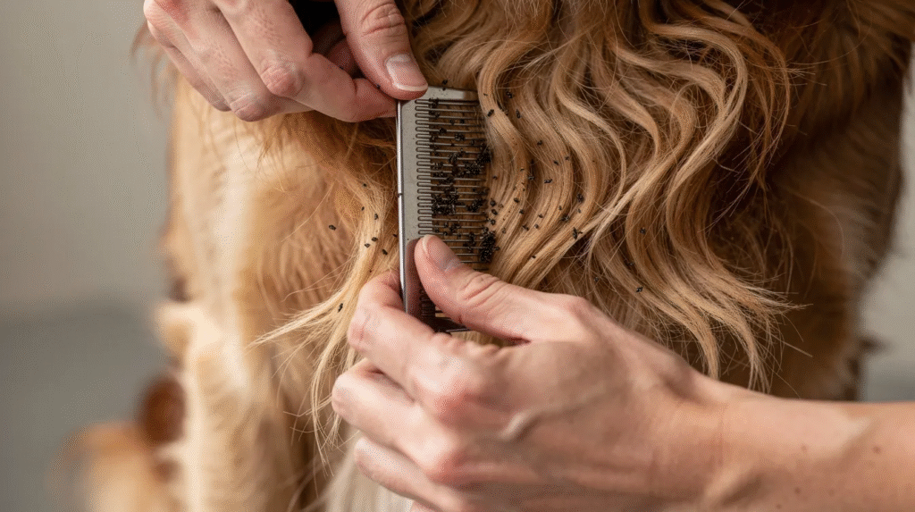
- Black flea dirt (digested blood) visible as tiny dark specks in the coat
- Scabs and excoriations from scratching
Mange patterns (sarcoptic and demodectic):
- Sarcoptic mange: Intense itch with crusty, thickened skin on ear margins, elbows, and hocks. Highly contagious between dogs. Mange causes intense itching and patchy hair loss that worsens rapidly without treatment.
- Demodectic mange: Patchy hair loss on the face and forelimbs, often in young or immunocompromised dogs, with less itch initially but risk of secondary infection.
High-resolution photos sometimes allow the AI to directly recognize fleas, ticks, or mite debris on the coat, which strengthens the confidence score. However, definitive mite diagnosis still requires skin scrapings or other tasks like PCR testing, so CompanAIn positions results as a suspicion level with clear recommendation to see a vet if mange is likely.
Fungal skin disease: ringworm and yeast dermatitis
Ringworm is a highly contagious fungal infection that affects dogs, cats, and humans-making it a genuine zoonotic risk for the whole household. Despite the name, it has nothing to do with worms.
Ringworm image features AI looks for:
- Circular or irregular bald patches with broken stubble hairs
- Fine scaling or crusty borders around the lesion
- Sometimes a pattern of central healing with an active, red outer ring
- Can appear anywhere but is common on the face, ears, and limbs
Yeast dermatitis presents differently:
- Greasy coat with a musty or “corn chip” odor
- Reddish-brown staining between toes or in skin folds (armpits, groin)
- Recurrent ear yeast infections with dark, waxy discharge
- Patterns AI can flag from photos of paws, armpits, and ears
AI can’t replace a fungal culture or PCR test, but it helps owners recognize when a “bald patch” or “smelly paw” could be a contagious or chronic fungal issue needing vet care. If ringworm is suspected, the report emphasizes the need for isolation and hygiene-washing bedding, disinfecting surfaces-to protect other pets and family members.
Bacterial skin infections (pyoderma) in dogs
Superficial pyoderma is a bacterial infection that commonly develops secondary to allergies, parasites, or skin trauma. It’s often mistaken for a “simple rash” but typically requires targeted treatment.
Visual signs AI looks for in image classification tasks:
- Red bumps (papules) and pustules with white or yellow centers. Bacterial skin infections appear as pus-filled blisters on the skin surface.
- Collarettes-circular rings of peeling skin left after a pustule ruptures
- Crusts, moist erosions, and patchy hair loss around the affected area
- AI can flag patterns associated with skin infections and distinguish allergic redness alone from redness plus pustules or collarettes that suggest infection detect requiring antibiotics or antiseptics
AI systems can assist in identifying skin infections in pets by catching these patterns early. Photos taken a few days apart can be compared by the app to show improvement or worsening, supporting decision-making with the vet about whether the current treatment plan is working.
Recognizing possible skin cancer in dogs from images
Most canine skin lumps are benign, but some are mast cell tumors, melanomas, or other forms of skin cancer that need early removal. AI can flag abnormal lesions but does not provide definitive diagnoses-biopsy and histopathology by board certified clinical pathologists remain essential.
Visual warning signs AI highlights:
- A rapidly growing lump that has changed noticeably over days to weeks
- Ulceration or bleeding on the surface of a mass
- Irregular borders or asymmetric shape
- Color changes-especially new dark skin lesions on lightly pigmented or hairless areas
- Basal cell carcinoma and other tumors that appear as firm, raised nodules
In a study of 664 canine masses, a non-invasive imaging tool achieved 85% sensitivity and 97% negative predictive value-meaning it was highly reliable at ruling out malignancy when it classified a mass as benign. This kind of risk stratification helps owners and vets prioritize which lumps need urgent biopsy.
Photograph new lumps with a coin or ruler for scale, then repeat images every one to two weeks. CompanAIn logs growth trends in the dog’s health timeline, and you can learn more about common benign masses in the guide to fatty lumps in dogs. Never delay vet care if the AI report mentions potential skin cancer risk.
Beyond the skin: ears, paws, nose, and other related checks
Many skin diseases in dogs start or become most visible in ears, paws, and other specialized skin regions that image analysis can assess alongside the coat.
Ear checks:
- Redness of the ear canal or inner pinna
- Brown or black waxy discharge (common in yeast or mite infestations)
- Yellow-green pus suggesting bacterial ear infections
- Crusty or thickened ear flap edges-sometimes linked to sarcoptic mange or vasculitis
Paw checks:
- Swelling between toes (interdigital cysts or furunculosis)
- Yeast staining and persistent licking patterns
- Crusts on paw pads, including hyperkeratosis
- Foreign-body tracks or draining tracts-the Pawgnosis model used object detection to identify pododermatitis and paw neoplasia from photos
Nose and lip checks:
- Crusting, depigmentation, or ulcers on the nose or lip margins
- These can be allergic, autoimmune, infectious, or neoplastic-AI flags them with clear recommendation to see a vet
CompanAIn’s broader body-part checks help link dermatological issues to systemic health concerns tracked elsewhere in the app, such as those flagged by the dog symptom checker.
What happens inside the algorithm: machine learning for dog skin images
CompanAIn uses deep learning architectures adapted from human dermoscopy research, modified for the unique challenges of fur coverage, breed variation, and the inconsistent lighting conditions of home photos. Feature extraction uses convolutional neural networks to analyze visual data, pulling out patterns invisible to the untrained eye.
Here’s what powers the system:
- Training inputs include millions of image patches annotated by veterinarians between 2019 and 2026, with labels for skin disease categories, severity, and body location. In human medicine, the ISIC challenge has over 80,000 labeled training images; veterinary datasets are growing rapidly along similar lines.
- The model first detects and segments the relevant skin area, then performs multi-label image classification-tagging combinations like “erythema,” “pustules,” and “alopecia” simultaneously. AI systems can classify multiple skin lesions with high accuracy using this approach.
- Pattern matching compares extracted features against labeled veterinary data to generate probability scores for each condition.
- Metadata such as age, breed, geographic region, and environmental factors can refine probabilities. For example, sarcoptic mange likelihood is weighted higher for dogs in crowded shelter environments than urban apartments.
- A 2026 study at Seoul National University trained EfficientNet models on canine skin images and achieved over 90% accuracy across all four lesion types tested-alopecia detection reached 98.1%.
In medicine, AI models trained on ISIC datasets have outperformed dermatologists by 11% in certain classification tasks. While veterinary datasets are still catching up, tools like VETSCAN IMAGYST from Zoetis already achieve accuracy comparable to expert pathologists when analyzing skin cytology samples, skin impression smears, ear swabs, and skin swabs. For more on how AI is transforming veterinary medicine broadly, see AI applications in veterinary medicine.

Accuracy, limitations, and how to interpret AI results
Independent clinical studies in human AI dermatology show accuracy comparable to dermatologists for many common skin diseases, and the veterinary field is following the same trajectory. AI identifies over 90% of common skin conditions in research settings, and a study demonstrated that multispectral imaging combined with standard photos achieved 87–89% accuracy for bacterial, fungal, and allergic skin disease classification in dogs. AI accuracy is comparable to dermatologists for common skin diseases in controlled study environments.
Practical limitations to keep in mind:
- Poor-quality images (blurry, overexposed, underexposed) degrade performance significantly
- Rare skin diseases are under-represented in training data-autoimmune conditions like pemphigus or unusual tumors may be missed
- Overlapping appearance between different rashes means some conditions look identical in photos
- Invisible internal causes (pain, fever, lymph node changes) can’t be assessed from a photo
- AI cannot replace critical laboratory tests required for confirmatory diagnosis, including skin scrapings, cultures, and histopathology
- AI systems require external validation before clinical deployment to ensure they perform across diverse breeds and coat types
When CompanAIn’s report shows multiple likely conditions, that uncertainty is a feature, not a flaw. AI outputs potential conditions with a confidence score during risk stratification, helping you understand which possibilities are most likely and which need ruling out. A “low confidence” result isn’t useless-it tells you the presentation is ambiguous and a vet visit is warranted.
Action guidance is phrased as “watch,” “contact your vet,” or “urgent visit” rather than definitive diagnoses. AI can improve the accuracy of veterinary diagnostics when used as a complement to professional judgment, not a substitute. Always share AI reports with your veterinarian through the secure vet portal for proper interpretation.
New insights from combining images with your dog's health data
Image analysis becomes a powerful tool when linked with timelines of symptoms, diet, environment, and lab results. AI can analyze pet health data for better diagnostics, and AI can provide personalized health insights for pet owners by connecting visual findings with the broader health picture.
Specific examples of what this combined approach reveals:
- Recognizing seasonal flares of atopic dermatitis from repeated spring photos-a pattern that might take years for an owner to notice unaided
- Correlating hair loss with abnormal thyroid labs flagged in your dog’s blood test results
- Linking belly rashes to a new detergent, bedding material, or diet change introduced weeks before the flare
- Spotting that ear infections cluster after swimming sessions or after a particular treat is introduced
CompanAIn’s dynamic health timeline shows clusters of dermatological events, allowing owners and vets to identify patterns and triggers over months or years. Anonymized data across thousands of dogs can also reveal population-level patterns-like a regional spike in parasite-related skin disease-feeding back into better AI performance over time.
How vets can use AI dermatology tools in daily practice
Veterinary professionals are key partners, not competitors, in AI-supported dermatology. Veterinary AI services can enhance vet-owner communication and care management across the board.
Practical in-clinic uses:
- Triaging skin cases from owner-submitted photos before appointments-teledermatology helps triage cases remotely through AI-assisted evaluations, saving valuable consultation time
- Documenting treatment response with serial images analyzed consistently by the AI model
- Educating owners with AI-generated visual reports, including heatmaps showing exactly where the lesion is most prominent
- Using the analysis to guide whether skin cytology samples, impression smears, or a send digital slide image referral is the appropriate next step
CompanAIn’s clinician-grade AI summaries and action plans save time in consultations by pre-organizing history, images, and suspected diagnoses. Vets can access a secure portal to review AI outputs, override or confirm suggestions, and add their own clinical notes. These corrections feed back into model refinement, improving future performance. Among zoetis offerings, the oclacitinib tablet (Apoquel) is frequently prescribed for allergic patients identified through these workflows-AI helps vets reach that decision point faster.
For a deeper look at how AI supports clinical decision-making, see AI tools for veterinary second opinions.
Ethical, privacy, and data-security considerations
Dermatological images of pets still involve owner privacy-backgrounds can reveal home environments, and metadata can contain location information. These images must be treated securely.
CompanAIn’s approach includes:
- Encrypted transmission of all images between the app and servers
- Storage in secure data centers with separation of image data from personally identifying information
- Strict access controls ensuring only authorized vets and owners can view specific records
- User control over whether images can be used in anonymized form to improve AI; clear opt-in options, never default enrollment
The system never uses images for advertising targeting. Anonymized datasets are focused exclusively on improving detection of skin diseases and skin cancer in dogs. AI models are validated as CE-marked Class I Medical Devices in Europe where applicable, reflecting a commitment to regulatory standards and clinical safety.
All research data used for training undergoes ethical review. Several published studies explicitly document owner consent and ethics committee approval for image use in model development.
When AI says "see a vet now": red-flag skin signs in dogs
Some skin presentations are emergencies or indicators of serious systemic disease. The AI model is programmed to escalate urgency when it detects these patterns.
Red flags that trigger immediate vet-care recommendations:
- Rapidly spreading bruised or purple skin (petechiae, ecchymoses)-may indicate a clotting disorder
- Extensive pustules combined with lethargy, fever, or loss of appetite
- Large, deep wounds, burns, or chemical exposure injuries
- Sudden widespread hives with facial swelling-possible severe allergic reactions or anaphylaxis
- Blackened or necrotic skin areas suggesting tissue death
- Bleeding skin lesions in older dogs, especially if ulcerated or rapidly growing
In these situations, CompanAIn’s report clearly advises emergency vet care. If you’re unsure whether your dog has a fever, the guide to checking for fever without a thermometer can help you act quickly at home while arranging transport.
Do not wait for AI results if your dog is collapsing, struggling to breathe, or in severe pain. Emergency care always overrides digital tools.
Using CompanAIn day-to-day for chronic skin conditions
Many dogs live for years with chronic skin disease-especially atopic dermatitis and recurrent infections-requiring ongoing monitoring rather than a one-time diagnosis and treatment cycle. AI tools help track changes in skin conditions over time between vet visits, turning reactive care into proactive management.
How owners can use the app on a regular basis:
- Log flare severity with photos weekly or monthly, noting the presence of redness, pustules, or hair loss in each image
- Mark new affected areas as they appear and compare with previous entries
- Track response to baths, diet changes, or new medications like an oclacitinib tablet or medicated shampoo
- Note environmental factors-season, humidity, pollen counts-alongside skin entries
AI trend detection can show whether a new treatment regime implemented in March 2025 actually reduced redness or hot-spot frequency over the following months. The severity index tracked across entries gives both you and your vet objective data to work with, rather than relying on memory alone.
Integration with reminders for parasite preventives, follow-up vet visits, and medication refills turns dermatology care into a structured, shared treatment plan between owner and vet. Early detection of health issues is crucial for proactive pet care management, and consistent tracking makes that detection possible.
How to talk to your vet about AI skin analysis results
Bring AI reports into the exam room as conversation starters, not conclusions. The goal is a collaborative conversation where AI reports support, not replace, veterinary expertise.
Practical tips:
- Share or print CompanAIn’s report before the appointment so your vet can review it alongside the medical record
- Highlight the time course shown in the health timeline-when the problem started, how it changed, and what you’ve tried
- Ask specific questions: “Could this be atopic dermatitis based on the pattern?” “Do we need a skin biopsy?” “Should we check thyroid levels?”
- Let your vet examine the dog and add their own findings-tactile information, lymph node palpation, and clinical judgment that no photo captures
Vets can use high-resolution images from the app in their medical record and to explain findings visually-showing you the difference between superficial infection and deeper skin disease. If the AI suggestion and vet impression disagree, resolve it in favor of the clinician while flagging the discrepancy to CompanAIn as valuable feedback that improves future model performance.
Future directions: where canine AI dermatology is heading
Between 2026 and 2030, canine AI dermatology will advance rapidly, driven by larger datasets, better smartphone cameras, and integration with wearable sensors. The research pipeline is full of promising results.
Emerging trends to watch:
- Multimodal models that combine images, text history, and lab data for higher diagnostic accuracy than any single input can achieve
- Real-time in-app lesion segmentation overlays that show owners exactly where lesion edges are and measure growth over time
- Prediction of flare risk in atopic dogs based on weather data, pollen counts, and regional parasite pressure
- Cross-species transfer learning from human skin disease models for rare canine conditions-while recognizing anatomical differences like fur and pigment distribution
- Non-visible spectrum imaging (infrared, multispectral, thermal) accessible through smartphone accessories, catching subclinical inflammation before it becomes visible
- Continued growth of labeled veterinary image databases, addressing current gaps in breed diversity, coat color representation, and dark skin pigmentation
The goal is earlier detection of serious issues like skin cancer and autoimmune skin disease in dogs, with AI acting as a continuous, at-home monitoring partner. AI will continue to complement, not replace, veterinarians in managing skin diseases-but it will make the partnership between pet owners and their vets more informed, more timely, and more effective.
Educational disclaimer and safe use of AI tools
CompanAIn’s image analysis is intended for educational and decision-support purposes only. It does not constitute a veterinary diagnosis or prescription. Acne vulgaris classification models from human dermatology, for instance, do not directly transfer to veterinary medicine-each species requires dedicated research and validation.
Key safety points:
- Always consult a licensed veterinarian for persistent, severe, or worrying symptoms
- Do not use AI tools to self-medicate with prescription drugs (antibiotics, steroids, immunosuppressants) without veterinary guidance
- In emergencies-collapse, difficulty breathing, severe pain-call your vet or local animal hospital immediately
- Limitations in datasets mean some rare skin disease patterns, including unusual presentations of skin cancer like certain melanomas, may not be recognized accurately
- AI identifies over 90% of common skin conditions in controlled settings, but real-world performance depends on photo quality, breed representation, and disease prevalence
Treat AI as a way to ask better questions and seek timely vet care-not as a replacement for professional examination by clinical pathologists or veterinary dermatologists.
Getting started with CompanAIn for your dog's skin health
Getting started takes less than five minutes and can immediately change how you monitor your dog’s skin.
Here’s the path for new users:
- Download the CompanAIn app and create a profile for your dog.
- Set your dog’s age, breed, weight, and any known allergies or ongoing medications.
- Perform an initial baseline skin check by uploading images of key areas: belly, ears, paws, flanks, and any current areas of concern.
- Enable sharing with your primary veterinarian through the secure vet portal so your patients-your pets-have continuous, connected care.
- Explore the dynamic health timeline to see how skin entries fit alongside other health data.
Both free and premium plans are available. The free plan includes a set number of skin image analyses per month, while the premium plan unlocks unlimited analyses, AI chat for follow-up questions, trend detection across your timeline, and clinician-grade summary reports you can share directly with your vet.
Proactive monitoring of skin conditions helps catch problems earlier, improves your dog’s daily comfort, and can extend healthy years of life. Whether it’s a mysterious rash, a recurring itch, or a new lump that appeared overnight, uploading that first photo is the simplest step you can take toward better skin health for your dog.